
|
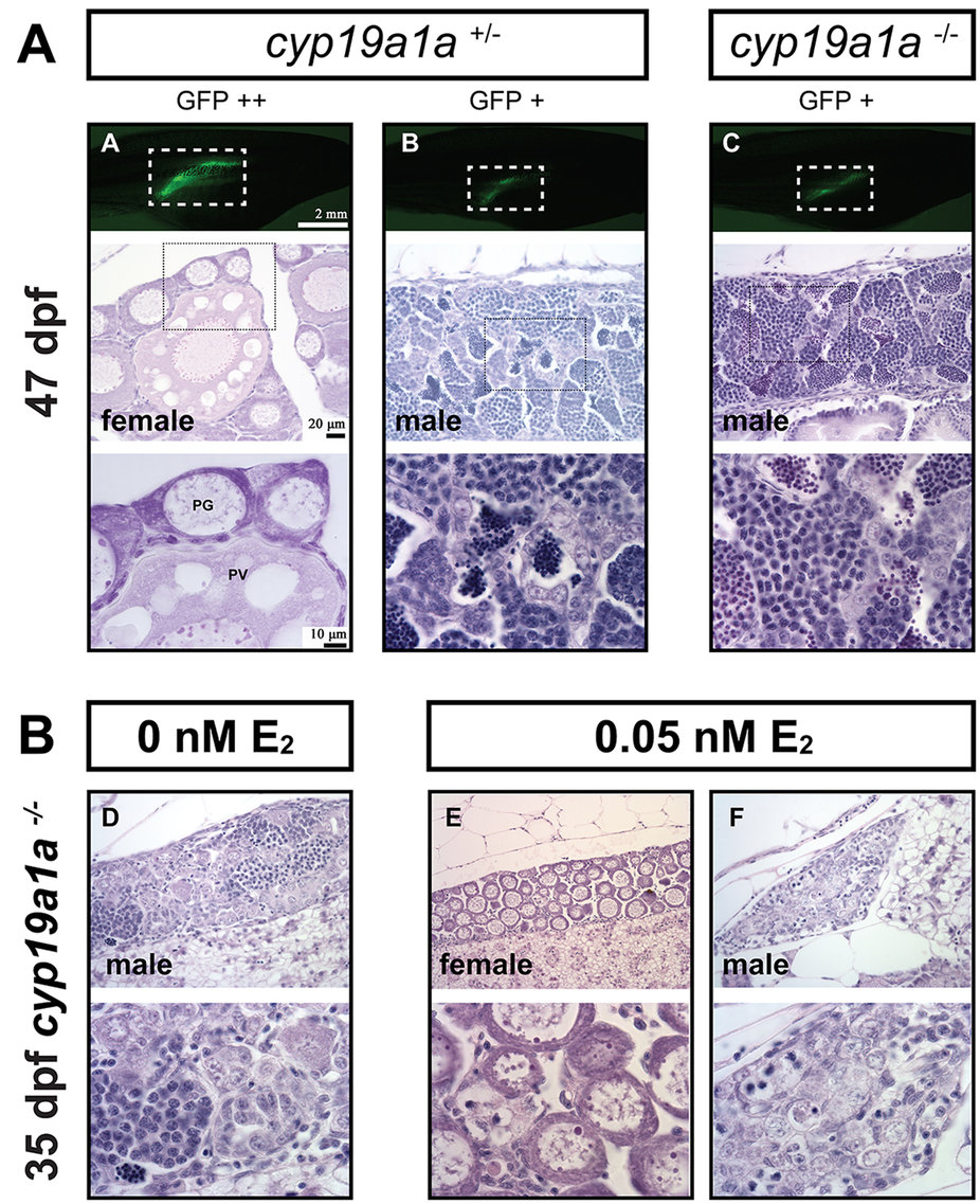
Fig. 8
(A) Gonad development at 47 dpf in the control (cyp19a1a+/−) and mutant (cyp19a1a−/−). The gonadal differentiation had finished in the control fish with the ovary and testis well developed. The ovary contained both PG and PV follicles, indicating puberty onset in the female. All mutant fish were males with well-developed testes containing all stages of spermatogenic cells without any oocytes. (B) Rescue of mutant phenotype by E2 treatment. The juvenile mutant fish were treated with E2 (0.05 nM) from 15 to 30 dpf, and the fish were sampled for histological examination at 35 dpf. E2 treatment could induce normal ovarian formation in some mutant fish as shown by fish E. Other fish (e.g., fish F) still had undifferentiated gonads that were likely destined to males.