
|
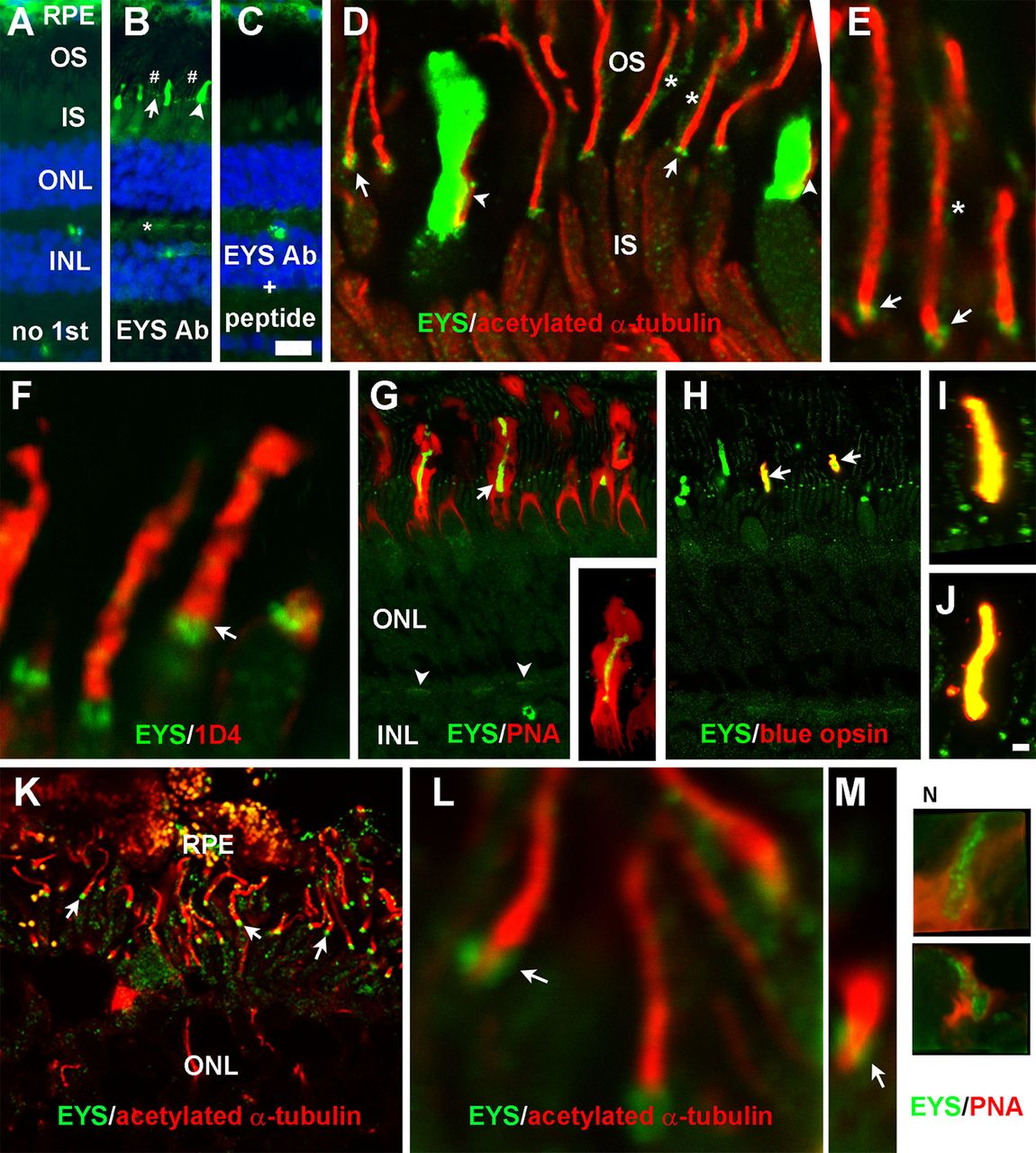
Fig. 2
EYS protein in primate retina was concentrated not only at the CC/TZ but also in cone outer segments and was weakly expressed at rod outer segments and cone terminals. Eye cups from three 8-year-old female rhesus macaque monkey and a human donor of undisclosed age and sex were stained with antibodies against EYS, acetylated α-tubulin, rhodopsin, or blue opsin and PNA. (A-C) Staining with no primary antibody, EYS antibody, and EYS antibody and blocking peptide, respectively. Note EYS immunoreactivity was observed as punctate pattern (arrow), cone outer segment (arrowhead), and rod outer segment (# signs) between the RPE and ONL as well as cone terminals (asterisk). (D,E) Double immunostaining of EYS (green) and acetylated α-tubulin (red). Note EYS reactivity was located on the basal end of acetylated tubulin (arrows), rod outer segment (asterisks), and adjacent to cone axonemes (arrowheads). (F) Double staining of EYS antibody (green) with rhodopsin antibody 1D4 (red). EYS is concentrated in the position basal to 1D4 immunoreactivity (arrow) and some of the EYS reactivity overlapped with 1D4. (G) Double staining of EYS antibody (green) with PNA (red). Note that EYS immunoreactivity in the cone outer segment was completely wrapped by PNA-stained cone matrix. EYS immunoreactivity in the OPL was also reactive to PNA (arrowheads). Insert in G shows a single cone at higher magnification. (H-J) Double staining of EYS (green) and blue opsin (red) antibodies. Note that a subset of EYS-positive cone outer segment was immunoreactive to blue opsin antibody (arrows) and there is a complete overlap between EYS and blue opsin reactivities (I,J). (K-M) Double staining of EYS (green) and acetylated α-tubulin (red) of human retina. Note that EYS immunoreactivity was located on the basal end of acetylated α-tubulin immunoreactivity (arrows). (N) Some human EYS immunoreactivity was located within PNA-positive domain. Scale bar in C: 20 µm for A-C; scale bar in J: 2 μm for E,F,I,J,L-N; 5 μm for D; 10 μm for K; 20 μm for G,H. IS, inner segment; INL, inner nuclear layer; ONL, outer nuclear layer; OS, outer segment; PNA, peanut agglutinin; RPE, retinal pigment epithelium.