
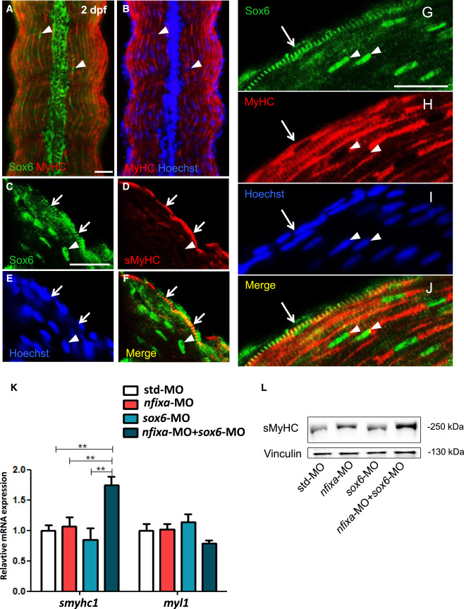
Fig. 6
|
Fig. 6
Functional Cooperation of Sox6 and Nfixa Is Conserved in Zebrafish
(A–J) Immunofluorescence with anti-Sox6 antibody (green) and anti-MyHC antibody (red) (A, B, and G–J) or with anti-Sox6 (green) and anti-sMyHC (F59, red) (C–F) on 2 dpf zebrafish muscle longitudinal sections. Arrowheads indicate Sox-positive nuclei in fast-twitch muscle fibers. Arrows indicate Sox6 staining in the cytoplasm of superficial slow fibers. Approximately one-fifth of the sMyHC-positive superficial cells displayed cytoplasmic Sox6 staining, whereas fast fibers negative for sMyHC only displayed nuclear Sox6 staining. Nuclei are counterstained with Hoechst. Scale bars, 25 μm.
(K) Quantitative real-time PCR analysis on trunk and tail regions at 48 hpf from embryos injected with std-MO or suboptimal doses of nfixa-MO (0.25 pmol), sox6-MO (0.1 pmol), or nfixa-MO + sox6-MO (∗∗p < 0.01; N = 2).
(L) Western blot for sMyHC at 52 hpf on trunk and tail regions of embryos injected with std-MO or suboptimal doses of nfixa-MO, sox6-MO, or nfixa-MO + sox6-MO. Vinculin was used to normalize the amount of loaded proteins.