
Image
|
Figure Caption
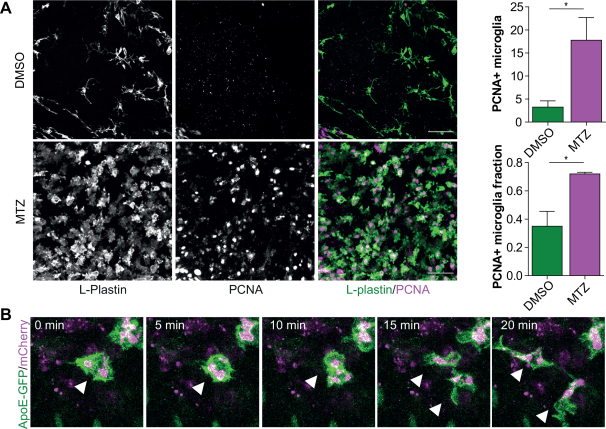
Fig. 5
Microglia proliferation upon the induction of neuronal cell death. (A) Immunofluorescence staining in the olfactory bulbs of 3-month-old treated MTZ-treated and DMSO-treated (control) fish. (B) Intravital imaging in 7 dpf zebrafish larvae with Apoe-driven GFP undergoing NTR-mediated neuronal cell death, showing the presence of dividing zebrafish microglia upon neuronal death. n = 3. Scale bar = 40 µm in (A). For quantification in (A) cells were counted in 3 selected volumes in the olfactory bulb (4.0 × 10 − 4 mm3) per fish (n = 3). Error bars represent standard deviation, *P < 0.05 (Student's t-test).
Acknowledgments
This image is the copyrighted work of the attributed author or publisher, and
ZFIN has permission only to display this image to its users.
Additional permissions should be obtained from the applicable author or publisher of the image.
Full text @ Glia