
|
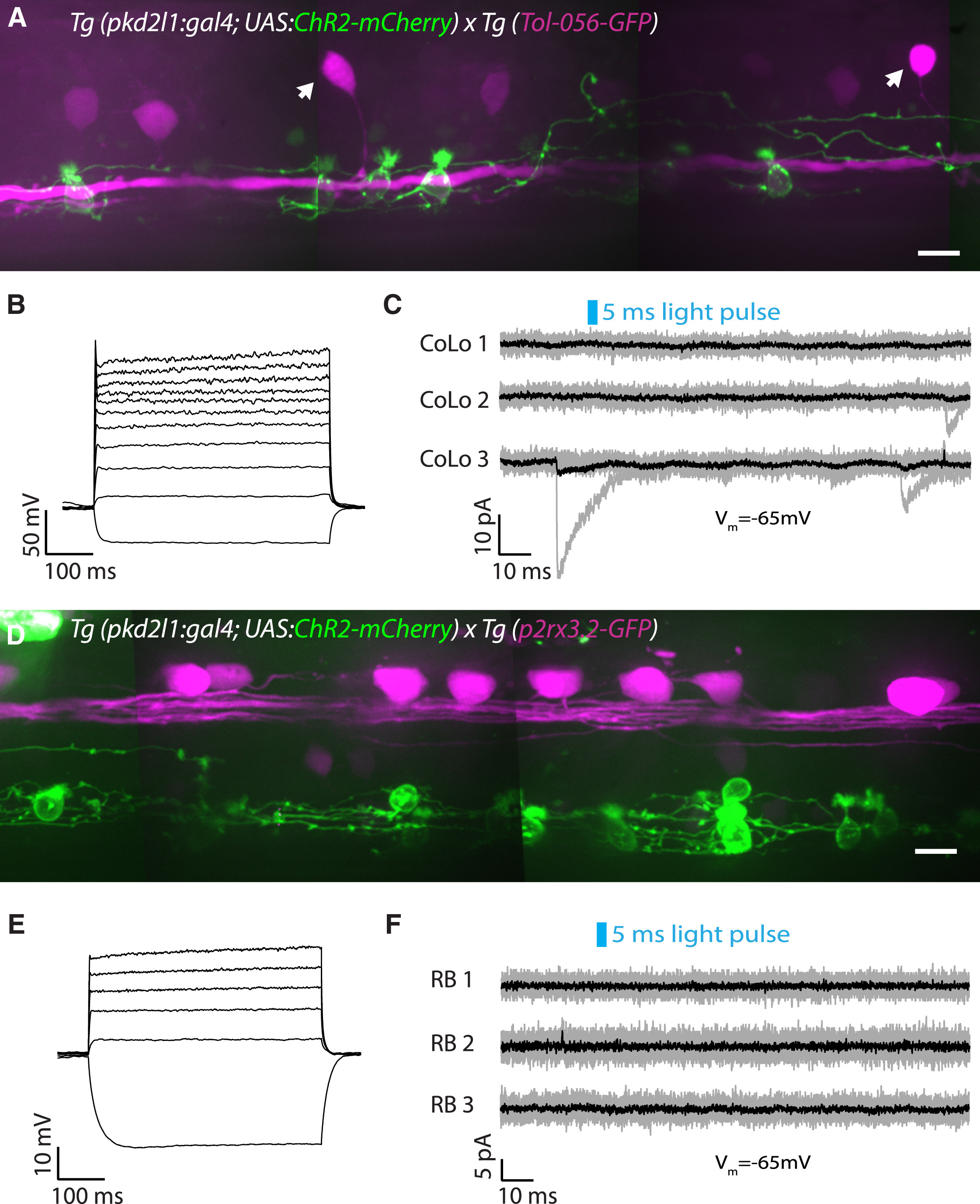
Fig. 5
CSF-cN Local Innervation onto the Escape Circuit Is Restricted to Excitatory Interneurons and Motor Neurons
(A) Z projection stack of CoLo glycinergic premotor interneurons expressing GFP (magenta) and CSF-cNs (green) in a Tg(pkd2l1:gal4; UAS:ChR2-mCherry; Tol-056-GFP) transgenic larva at 3 dpf. Arrows indicate CoLo cell bodies. Scale bar, 10 μm.
(B) Current-clamp recording showing the typical firing pattern of a CoLo neuron with a single weak action potential in response to current injection (steps of 20 pA from −30 pA to +370 pA).
(C) CSF-cN stimulation elicited by a 5-ms light pulse fails to induce an IPSC in CoLos. Example of voltage-clamp recordings from three CoLos (Vm = −65 mV) showing no IPSCs following 5-ms light pulses (black trace is the average of ten trials shown in gray). IPSCs in CoLos were never observed following CSF-cN stimulation (n = 13 cells).
(D) Z projection stack showing Rohon-Beard neurons expressing GFP (magenta) and CSF-cNs (green) in a Tg(pkd2l1:gal4; UAS:ChR2-mCherry; p2rx3.2:GFP) transgenic larva at 3 dpf. Note that the axonal projections of CSF-cNs do not reach Rohon-Beard somas or axons. Scale bar, 10 μm.
(E) Current-clamp recording showing the typical firing pattern of a Rohon-Beard neuron with a single weak action potential in response to current injection (steps of 20 pA from −30 pA to +170 pA).
(F) CSF-cN stimulation elicited by a 5-ms light pulse fails to induce an IPSC in Rohon-Beard (RB) neurons. Example of voltage-clamp recordings from three Rohon-Beard neurons (Vm = −65 mV) showing no IPSCs following 5-ms light pulses (black trace is the average of ten trials shown in gray). IPSCs in Rohon-Beard neurons were never observed following CSF-cN stimulation (n = 10 cells).
See also Table S1.