
|
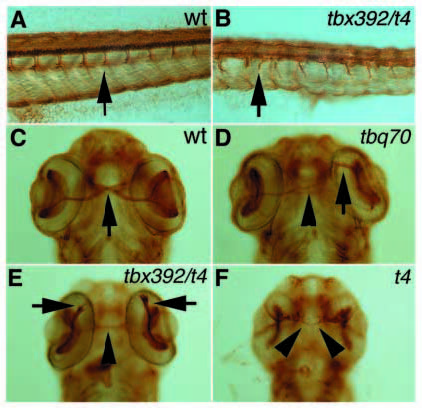
Fig. 6
Zn5 antibody staining of secondary motorneurons in a wildtype (A) and a syutbx392/syut4 mutant (B) embryo at 52 hpf. In syu mutant embryos the axons of the secondary motorneurons (indicated by arrows) fail to branch and instead cease to extend or grow further ventrally in an abnormal pattern. Zn5 staining of retinal axon projections in wild-type (C) and syu mutant alleles (D-F) at 52 hpf. In wild-type embryos, the retinal ganglion cell axons have crossed the midline where the optic chiasm is formed (arrow) and grow towards the tectum on the contralateral side. In syu mutant embryos, these retinal axons frequently fail to cross the midline and remain on the ipsilateral side (arrows) or, if they do cross the midline, frequently grow abnormally and in many cases only form very thin axon bundles (arrowheads).