
|
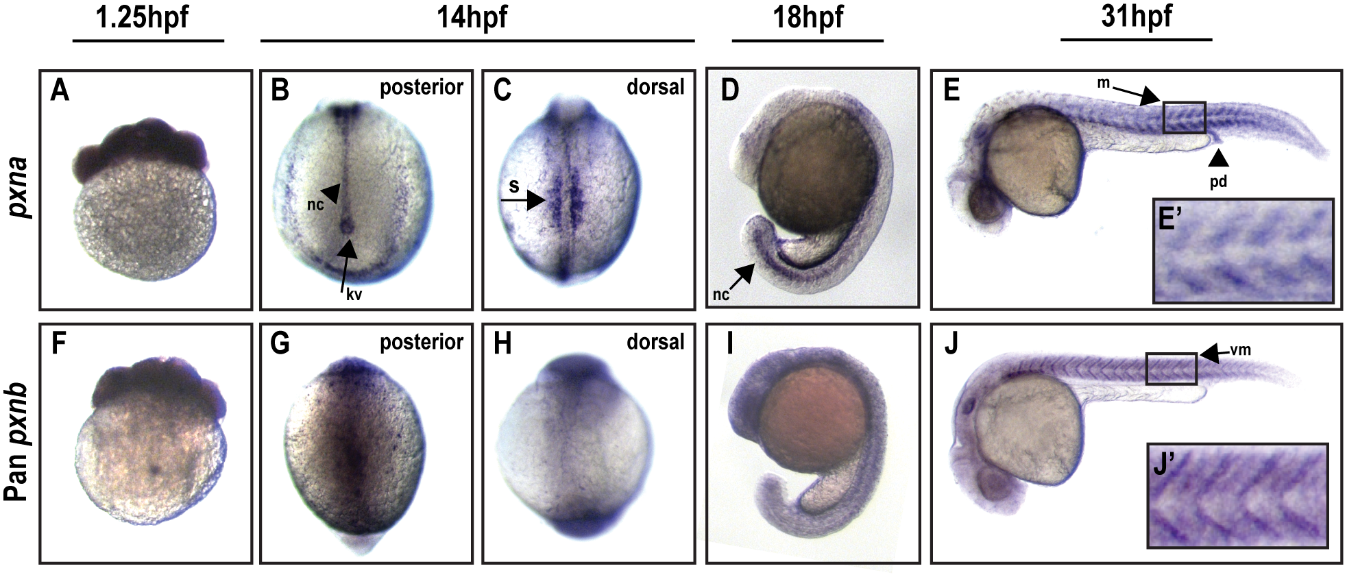
Fig. 6
Spatial expression of Paxillin orthologs during early zebrafish development.
(A-E) RNA in situ hybridizations using a pxna antisense probe detected maternal transcripts at 1.25 hpf (A) and enriched expression in posterior notochord (nc), Kupffer’s vesicle (kv), and somites (s) at 14 and 18 hpf (B-D). At 31 hpf (E), pxna expression was detected in pronephric ducts (pd) and myotomes (m). E’ depicts zoomed-in boxed regions of E. (F-J) A pan-pxnb antisense probe detected maternal mRNA (F) and unrestricted expression at 14 and 18 hpf (G-I). pxnb expression was detected in myotomes and vertical myosepta (vm) at 31 hpf (J). J’ depicts zoomed-in boxed regions of J.