
|
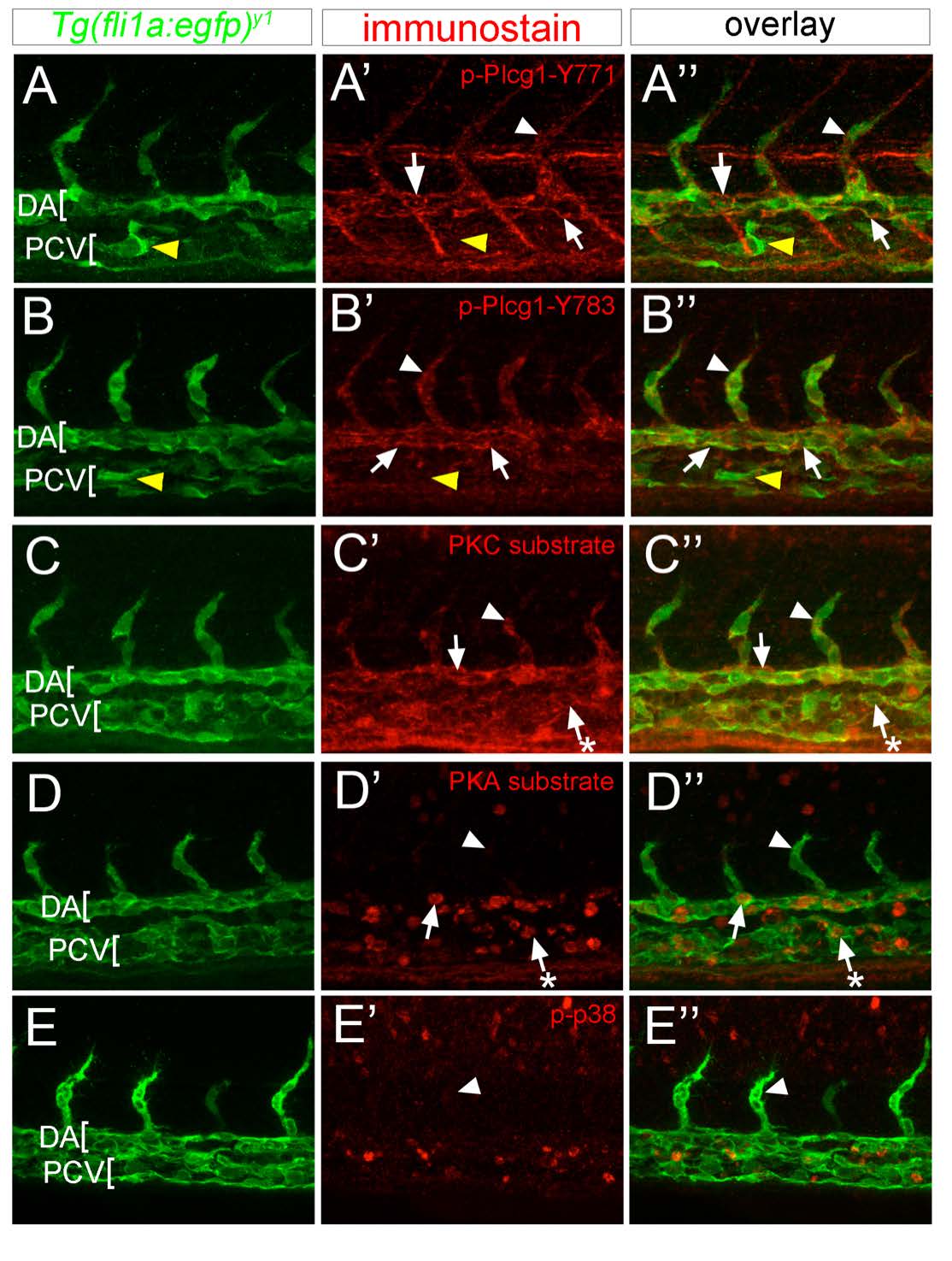
Fig. S1
Activation of presumptive Vegfa downstream signaling effectors in zebrafish blood vessels. (A-E) Confocal images of wild type Tg(fli1a:egfp)y1 embryos at 24 hours post fertilization (hpf). Lateral views, anterior to the left, dorsal is up. Dorsal aorta (DA) and posterior cardinal vein (PCV) are indicated. Arrowheads indicate intersomitic vessels, arrows indicate DA endothelial cells, and yellow arrowheads denote PCV endothelial cells. (A'-E') Images from same embryos as in A-E, immuonstained with primary antibody against indicated epitope and secondary antibody conjugated to HRP followed by TSA-Cy3 amplification. (A''- E'') Overlay of images from Tg(fli1a:egfp)y1 expression and Cy3 fluorescence.