
|
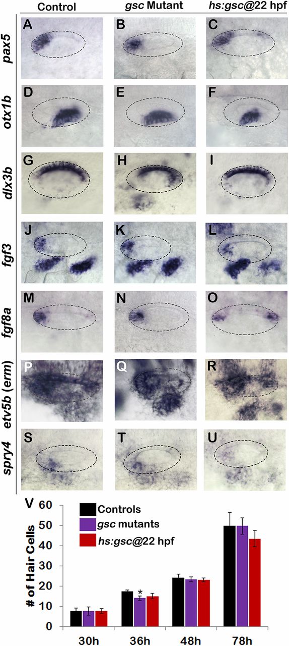
Fig. S3
Effects of altering gsc function on otic vesicle patterning. (A–U) Whole-mount images (dorsal up, anterior left) show dorsolateral views of the otic vesicle (outlined) in controls, gsc mutants, and hs:gsc embryos for the indicated genes at 24 hpf. Patterning of the otic vesicle is not affected in gsc mutants. Hs:gsc embryos show a slight decrease in the expression of ventral-lateral otic marker otx1 (F) but otherwise appear normal. (V) Means and SD of the total number of hair cells in utricular and saccular maculae of control embryos, gsc mutants, and hs:gsc embryos at the indicated times (n = 3 embryos each). Significant differences (P < 0.05) from the control are indicated by asterisks. Data were obtained by counting hair cells in the serial sections. Accumulation of the hair cells was normal except that gsc mutants showed a small but significant decrease relative to the control at 36 hpf. There was also a slight decrease in the number of hair cells (though not statistically significant) in hs:gsc embryos at 78 hpf.