
|
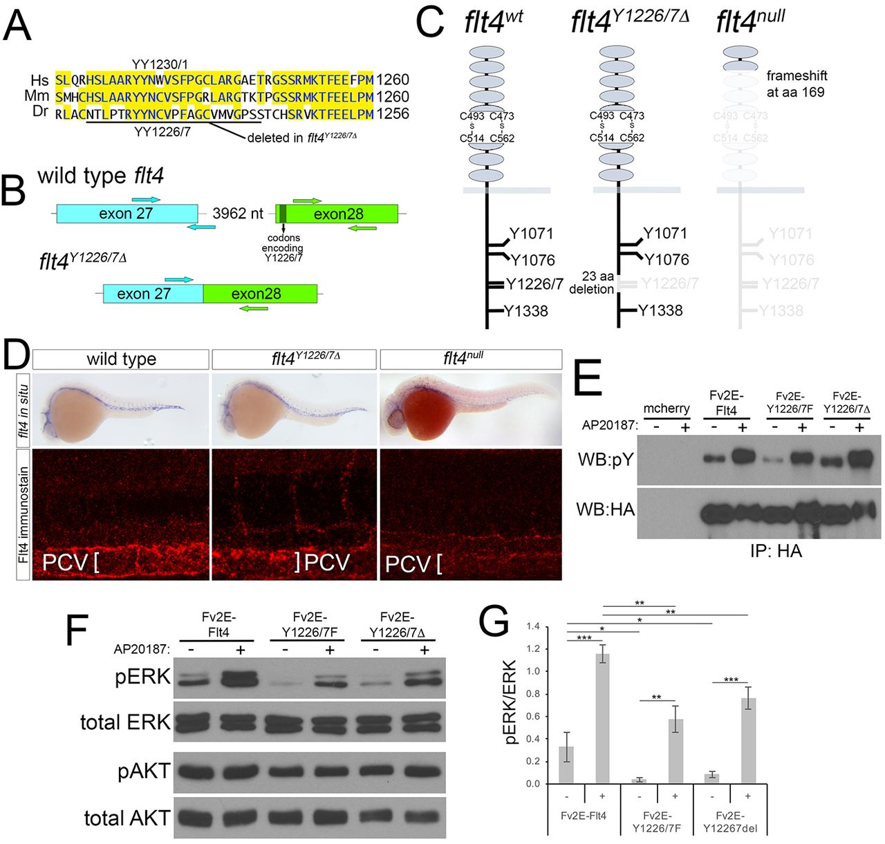
Fig. 1
Targeted deletion of Flt4 Y1226/7. (A) Flt4 cytoplasmic domains from human (Hs), mouse (Mm), and zebrafish (Dr). YY1226/7 and 23 amino acid deletion in flt4Y1226/7Δ mutants are shown. (B) Schematic of wild-type flt4 and flt4Y1226/7Δ genomic loci. Positions of TALEN pairs in exon 27 and exon 28 are indicated by arrows and codons encoding Y1226/7 are noted. (C) Schematics of wild-type, Flt4Y1226/7Δ and Flt4null receptors. Residues eliminated by targeted deletions are grayed out. (D) Top: whole-mount in situ hybridization using a digoxigenin-labeled antisense flt4 riboprobe on embryos of the indicated genotype at 25 hpf. Bottom: whole-mount immunostaining using a polyclonal antibody against zebrafish Flt4 at 30 hpf. (E) Immunoprecipitates from human embryonic kidney cells transfected with the indicated constructs and treated with AP20187 to dimerize Fv2E (+), or left untreated (−). Antibodies used for immunoprecipitation (IP) or western blot are indicated. pY, phophotyrosine; HA, hemagglutinin. (F) Western blot analysis of lysates from cells transfected with the indicated construct and treated with AP20187 (+), or left untreated (−). (G) Quantification of pERK/ERK ratio from triplicate western blots (as in F). *P<0.05, **P<0.01, ***P<0.001; error bars are ±s.d.