
Fig. S3
- ID
- ZDB-IMAGE-161130-8
- Publication
- Grainger et al., 2016 - Wnt9a Is Required for the Aortic Amplification of Nascent Hematopoietic Stem Cells
- All Figures
- Figures for Grainger et al., 2016
|
Fig. S3
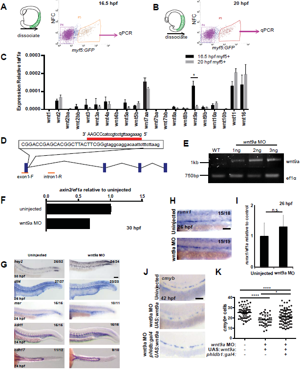
qPCR screen results and wnt9a morpholino validation.
GFP+ cells were sorted by FACS from 16.5 (A, C) and 20 (B, C) hpf myf5:eGFP embryos (n=200 at each time point), RNA isolated and qPCR performed for Wnt genes. Knockdown of wnt9a was achieved with a MO (D). Intron retention was verified by PCR (n=10 at each dosage) (E). F. Loss of canonical Wnt activity was verified by qPCR for axin2 on whole embryos (n=30) at 30 hpf. G. The wnt9a MO did not affect formation of the aorta (30 hpf hey2; 26 hpf dll4) vasculature (26 hpf msr; 24 hpf flk,), or pronephros (24 hpf cdh17). Examination of HSPC marker runx1 at 26 hpf by WISH (H) and qPCR (n=20) (I) in wnt9a morphants, compared to uninjected controls. J. Examination of HSPC marker cmyb at 42 hpf by WISH in wnt9a morphants in the presence or absence of somitic wnt9a, compared to uninjected controls. K. Quantification of cmyb+ cells from J. Scale bars=0.1 mm. *P<0.05, ****P<0.0001, n.s.=not significant. Error bars represent standard deviation.