
|
Fig. S2
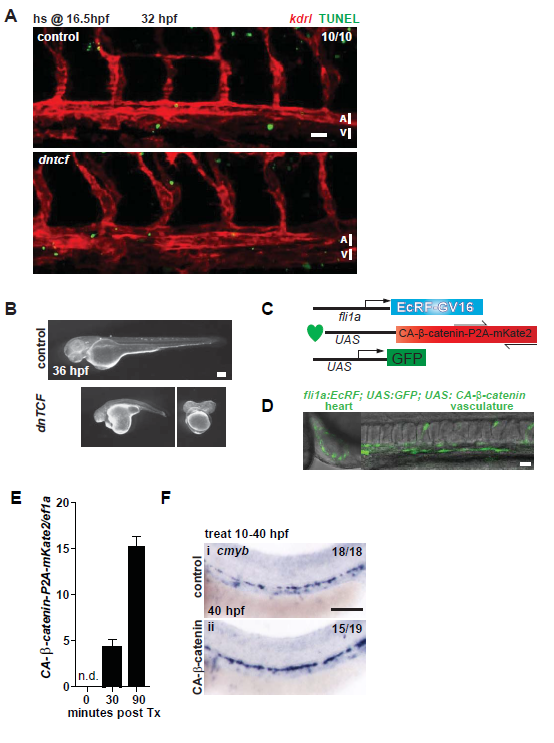
Apoptosis in the aorta and validation of UAS:dntcf and UAS:CA-β-catenin lines.
A. hs:dntcf; kdrl:mCherry fish were examined for apoptosis by TUNEL assay at 32 hpf after a 10 minute heat shock at 16.5 hpf. Scale bar= 30µm. B. The UAS:dntcf line was validated by crossing it to hsp:Gal4 fish and heat shocking at 38°C for 30 minutes at 13 hpf, which results in severe axial truncations, shown at 36 hpf. Scale bar= 100µm. C. To assess the effect of Wnt activation in the endothelium on HSC development, we generated a transgenic zebrafish line (UAS:CA-β-catenin) carrying a constitutively active form of β-catenin (CA-β-catenin) under the control of a drug inducible, endothelial specific Gal4 driver (fli1a-EcR), where Gal4 is shuttled to the nucleus after Tebufenozide (Swift et al., 2014). D. Activation of the transgenes (UAS:CA-β-catenin; fli1a-EcR; UAS:eGFP) was monitored by GFP expression in the heart and vasculature. Scale bar= 30µm E. Expression of CA-β-catenin was detectable 30 minutes after treatment by reverse transcription quantitative PCR (qPCR). F. UAS:CA-β-catenin; fli1a- EcR fish were analyzed at 40 hpf by WISH for cmyb after treatment from 10-40 hpf with 0.2µM Tebufenozide. Scale bar= 0.1mm.