
|
Fig. 2
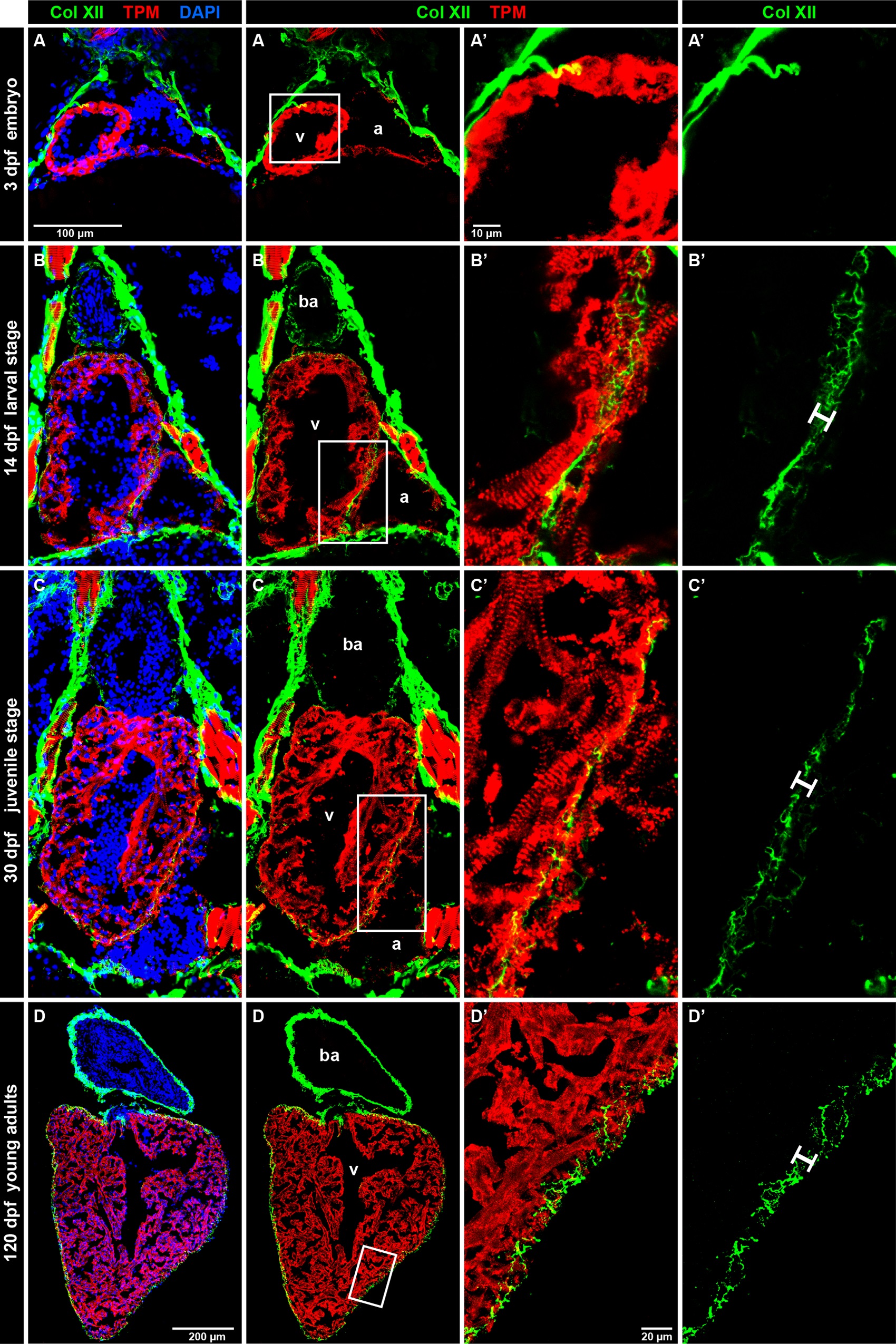
Developmental dynamics of Col XII expression in the zebrafish heart.
(A-D) Longtudinal sections of the zebrafish heart after double immunostaining against Col XII (green) and Tropomyosin (red), with DAPI contrastain (blue). dpf, days post-fertilization. Three chambers of the zebrafish heart: v, ventricle; a atrium; b.a., bulbus arteriosus (non-muscular structure). N ≥ 5. (A) At 3 dpf, embryos express Col XII in the pericardium, but not in the heart. The pericardial fibers seem to invade the surface of the heart. (B) At 14 dpf, the three chambers of the larval heart are surrounded by Col XII-positive fibrils within 10 μm of outer myocardial layer (white bar). (C) At 30 dpf, the juvenile fish heart contains a thickened myocardium (red), but the size of Col XII-positive layer remains unaltered. (D) At 120 days post fertilization, young adult fish maintain Col XII-labeled fibers along the heart circumference in a pattern similar to the one seen at the larval stage.