
|
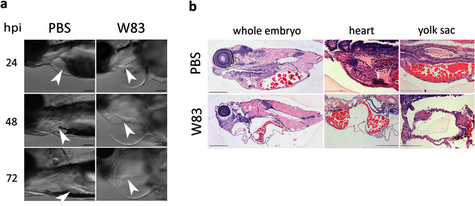
Fig. 2
Effect of wild-type Pg W83 on zebrafish larvae tissue structure.
Lateral view of impaired, elongated heart morphology of Pg W83-infected larvae; white arrowhead indicates heart. Heart chambers were distant and much smaller in comparison to PBS-injected control larvae (a). Sagittal histological sections of H&E stained 48 hpi Pg W83-infected larvae revealing advanced tissue damage in the cranial and cardiac regions along with yolk sac oedemas (b). In all figures larvae were infected with 5 × 10⁴ CFU Pg at 30 hpf W83 or PBS as a control. At least three individual experiments were performed and images are representative of at least n = 5 larvae per group in each experiment. Scale bars = 100 μm in (a) and 200 μm in (b).