
Image
|
Figure Caption
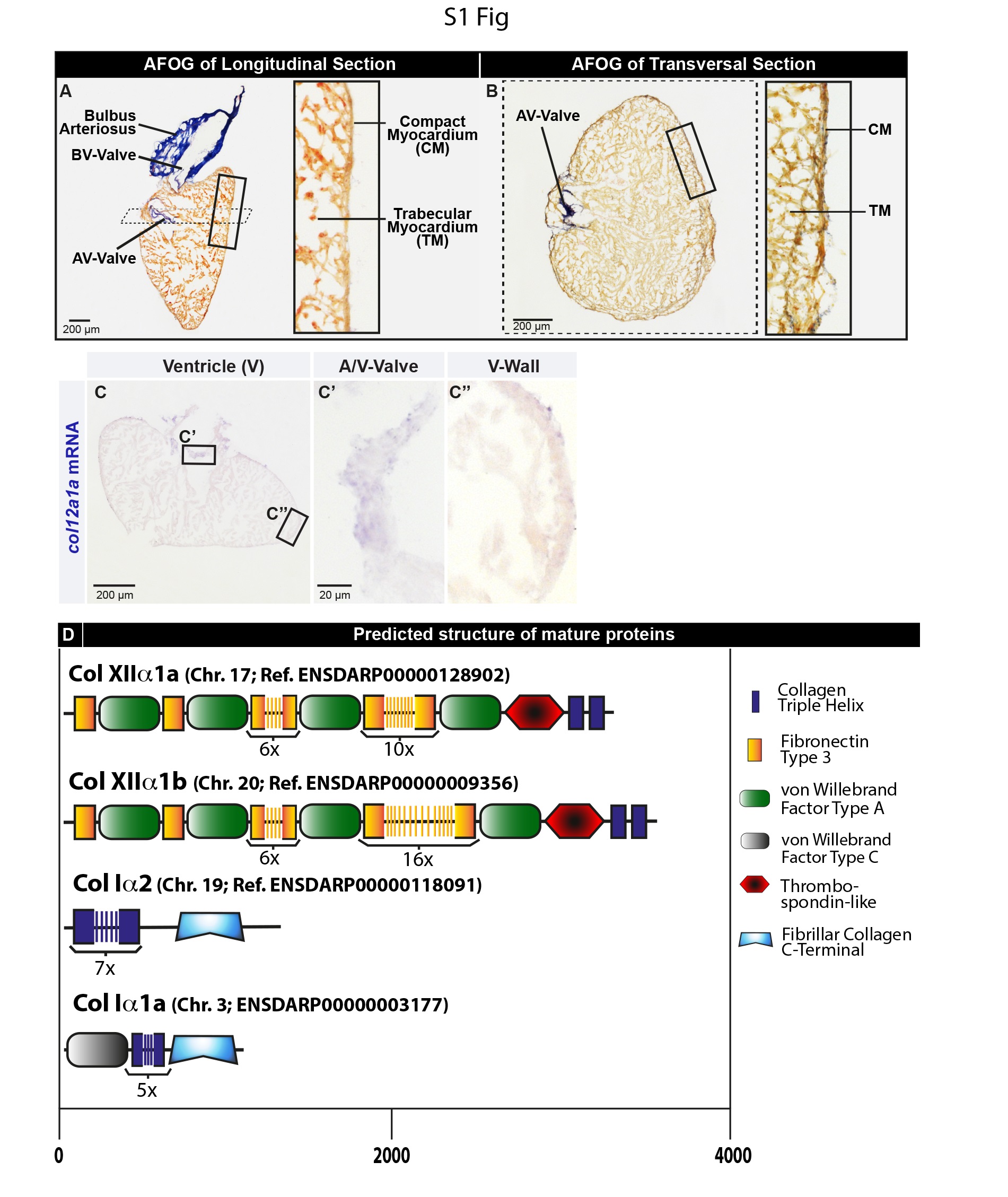
Fig. S1
The adult zebrafish heart and its collagenous components.
(A-B) Aniline blue, acid Fuchs in and Orange G (AFOG) histological staining demarcates the muscle of the ventricle (orange) and non-muscular structmes that are rich in collagen (blue). (A) The longitudinal section of the zebrafish heart shows a pyramidal shape of the ventricle and peer-shaped bulbus arteriosus of the outflow tract. The atrium is not included in the section, but the atria-ventricular valve is visible. (B) Transversal section of the zebrafish ventricle at the level of the atrio-ventricular valve. Magnified images of the ventricular wall (framed areas) reveal an outer layer ofa compact myocardium, which surrounds the main spongy (trabecular) myocardium. Little collagen (blue) is visible in the epicardium. (C) In-situ hybridization of tJ1e heart ventricle display a weak expression of coll 2a I a in tJ1e uninjured zebra fish heart. (D) Predicted mature protein structures ofzebrafish FACIT collagens: Collagen XII alpha la (Col XIlala) and Collagen XII alpha la (Col XII a I b); and fibril-forming collagens: Collagen I alpha I a (Col Ia I a) and Collagen I alpha 2 (Col la2), based on the conserved domain analysis of the annotated genes in the public database. Pre-pro-peptide sequences are not shown.
Acknowledgments
This image is the copyrighted work of the attributed author or publisher, and
ZFIN has permission only to display this image to its users.
Additional permissions should be obtained from the applicable author or publisher of the image.
Full text @ PLoS One