
Image
|
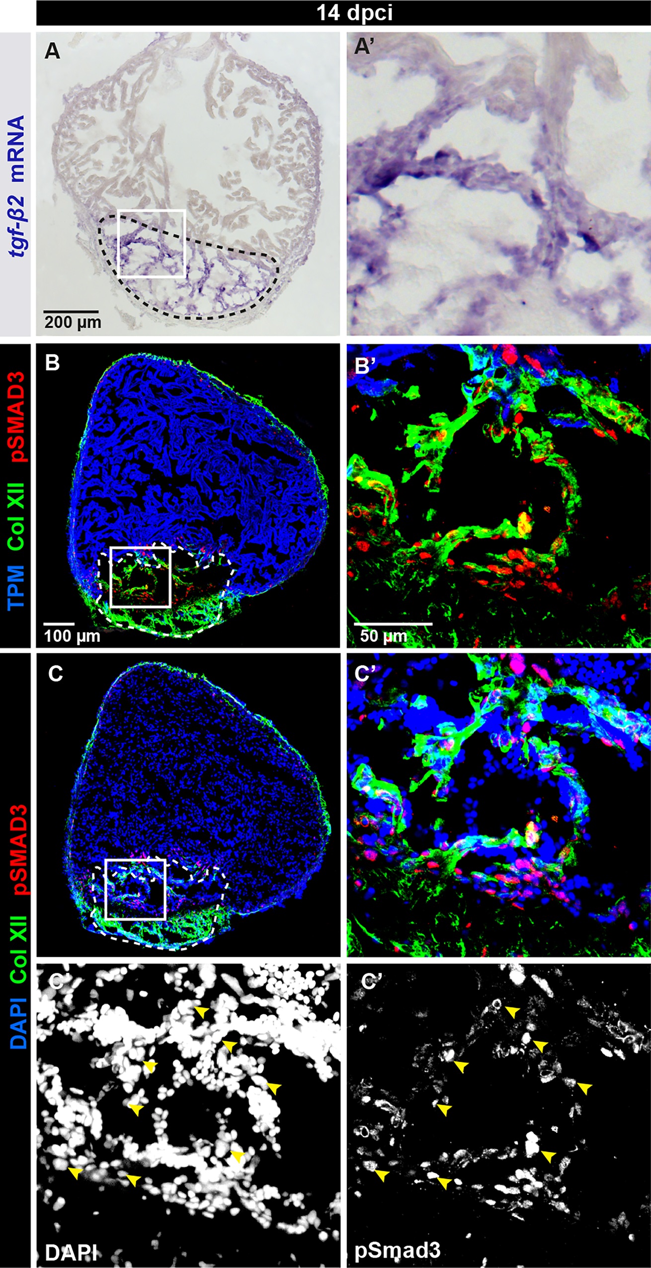
Figure Caption
Fig. 6
TGF-β signaling is activated in Collagen XII-positive fibrotic tissue.
(A-C) Analyses of ventricle sections at 14 dpci. (A) In -situ hybridization detects tgf-β2 expression in the cryoinjury area. N = 5. (B, C) Immunofluorescence staining reveals co-distribution of Col XII fibrils (green) and p-Smad3 (red) in the post-cryolesion zone that is recognized by the absence of Tropomyosin (blue in B). p-Smad3 reactivity is detected in the nuclei visualized by DAPI (blue in C). N = 5.
Acknowledgments
This image is the copyrighted work of the attributed author or publisher, and
ZFIN has permission only to display this image to its users.
Additional permissions should be obtained from the applicable author or publisher of the image.
Full text @ PLoS One