
|
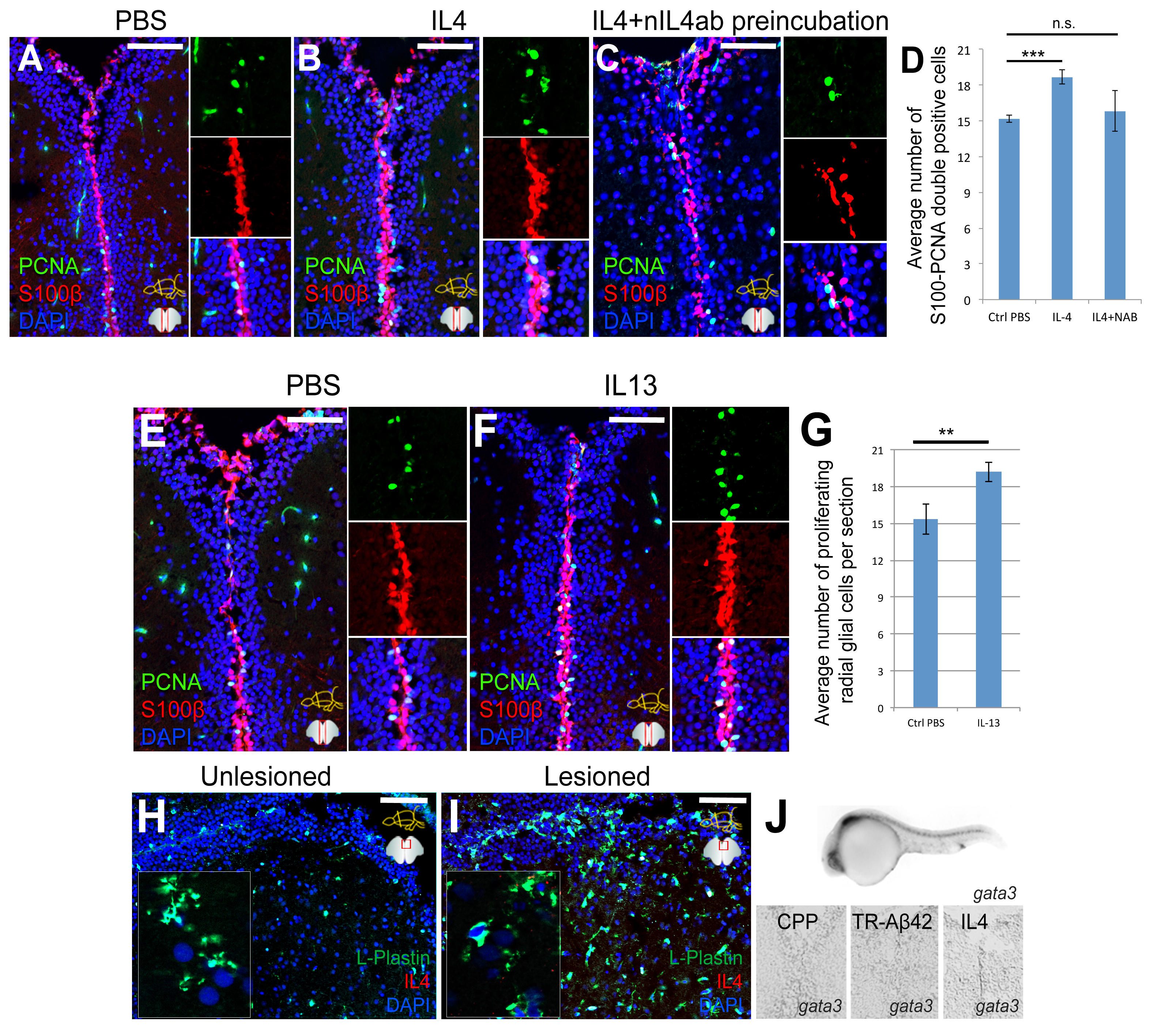
Fig. S4
Specificity of the IL4 neutralization, and the effects of IL13 on radial glial cell proliferation. Related to Figure 3, Figure 4.
(A-C) Immunohistochemistry (IHC) for S100beta and PCNA on fish brain injected with PBS (A), IL4 (B) and IL4 preincubated with the neutralizing antibody (C). Insets on the right of every panel show individual fluorescence channels and the composite from a close-up region.
(D) Quantification graph for A-C. Neutralizing antibody abrogates the inductive effect of IL4 on radial glial cell proliferation.
(E,F) IHC for S100beta and OCNA on fish brains injected with PBS (E) or IL13 (F). Insets on the right of every panel show individual fluorescence channels and the composite from a close-up region.
(G) Quantification graph for E and F. IL13 does not increase radial glial cell proliferation in adult zebrafish brain.
(H) L-Plastin and IL4 staining in unlesioned zebrafish brain. Inset is close-up image.
(I) L-Plastin and IL4 staining in zebrafish brain 3 days after lesion. Inset is close-up image.
(J) gata3 in situ hybridization in 24 hour post fertilization embryo, and in adult zebrafish brains injected with TR cell penetrating peptide (CPP), TR-Aβ42, and IL4. gata3 is expressed in embryos but not in adult zebrafish brain after TR-Aβ42 or IL4.
Scale bars 100 μm. Data are represented as mean ± SD.