
|
Fig. 1
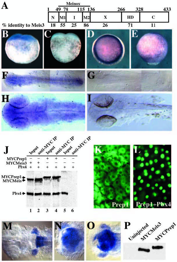
Prep1 retains functions similar to Meis3. (A) Prep1 protein. Letters indicate the name of individual domains; the Meinox domain includes the M1, I and M2 domains. Numbers on top represent amino acid positions in Prep1 and numbers on the bottom indicate percent identity of each domain between Prep1 and Meis3. (B-I) Expression pattern of prep1 during zebrafish embryogenesis. An antisense (B,D,E,F,H) or sense (C,G,I) probe for prep1 was hybridized to zebrafish embryos at the two-cell stage (1 hpf; B,C), early gastrula (6 hpf; D), late gastrula (9 hpf; E), early segmentation (13 hpf; F,G) and late segmentation (25 hpf; H,I). (B,C) Lateral views with animal pole towards the top. (D) An animal pole view. (E) A lateral view with dorsal towards the right and anterior towards the top. (F-I) Dorsal views with anterior towards the left. (J) Prep1 binds to Pbx4/Lzr in vitro. Pbx4/Lzr was in vitro transcribed in the presence of 35S-methionine together with MycMeis3 (lanes 1, 2), MycPrep1 (lanes 3, 4) or by itself (lanes 5, 6), immunoprecipitated with anti-Myc antibody, resolved on a 10% SDS-PAGE gel and exposed to film. (K,L) Prep1 is brought to the nucleus by Pbx4/Lzr. One- to two-cell stage embryos were injected with 300 pg MycPrep1 mRNA by itself (K) or together with 300 pg pbx4/lzr mRNA (L), raised to 5 hpf and stained with anti- Myc antibody. (M-O) Prep1 induces hindbrain fates in the same way as Meis3. One- to two-cell stage embryos were injected with 500 pg lacZ RNA (M), meis3+pbx4+hoxb1b mRNA (N; 165 pg each), or prep1+pbx4+hoxb1b mRNA (O; 165 pg each), raised to 25 hpf and analyzed for hoxb2 expression by in situ hybridization. All three embryos are dorsal views with anterior to the left. (P) MycMeis3 and MycPrep1 are expressed at similar levels. One- to two-cell stage embryos were injected with 300 pg MycMeis3 mRNA or MycPrep1 mRNA, raised to 5 hpf, lysed, resolved on a 10% SDS-PAGE gel, western blotted and probed with anti- Myc antibody.