
Image
|
Figure Caption
Fig. 6
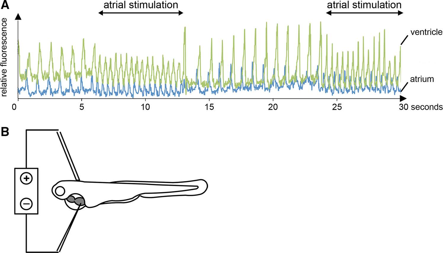
a Selective atrial stimulation of ste−/− mutants at 72 hpf reconstitutes heart rate to normofrequency as depicted by simultaneous measurements of atrial (blue) and ventricular (green) cytoplasmic calcium transients. b Schematic description of the experimental setup for selective atrial stimulation using an electric stimulator
Figure Data
Acknowledgments
This image is the copyrighted work of the attributed author or publisher, and
ZFIN has permission only to display this image to its users.
Additional permissions should be obtained from the applicable author or publisher of the image.
Full text @ Basic Res. Cardiol.