
|
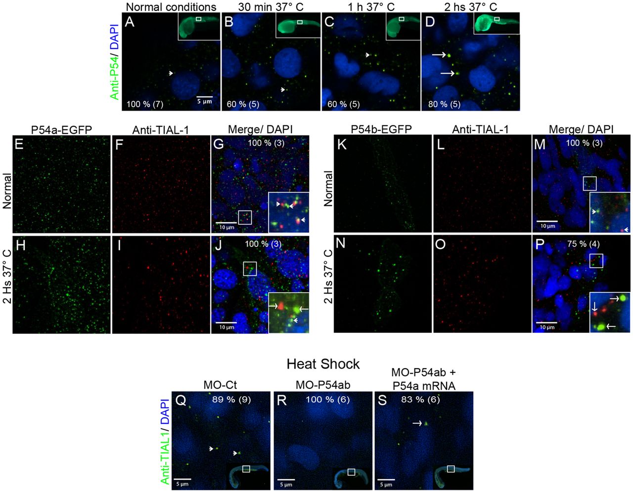
Fig. 3
Heat-shock induced the association of P54a and P54b RNA helicases with large cytoplasmic granules. (A,E–G,K–M,Q) Wild-type 24 hpf zebrafish embryos were maintained under normal conditions at 28.5°C or (B–D,H–J,N–P,R,S) under heat-shock conditions at 37°C. (A–D) Anti-P54 immunostaining in embryos subjected to heat-shock for 30 min, 1 h and 2 h. Nuclei are counterstained with DAPI. The top right insets are general views, and the amplified regions are indicated in white boxes. (E–J) Expression of the P54a-EGFP in combination with TIAL-1 immunostaining (as a stress granule marker). In the inset, some P54a-expressing granules co-localize with or are in close contact with TIAL-1-positive granules. (K–P) P54b-EGFP expression in small (at 28.5°C) or large (at 37°C) granules, combined with immunostaining with anti-TIAL-1. The insets show both P54b-expressing granules and TIAL-1-positive granules. Arrowheads point to small granules, such as P-bodies, and arrows indicate larger granules, such as TIAL-1-positive (red) stress granules. (Q–S) Knockdown of P54a and P54b expression by splice-blocking morpholinos prevents the formation of stress granules labeled by anti-TIAL-1. For each panel, the percentage of embryos showing the same pattern is indicated, and the total number of fish embryos tested is shown in parentheses. White arrowheads point to small granules (putative P-bodies) and white arrows to large granules (putative stress granules).