
|
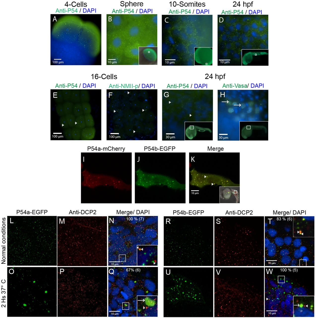
Fig. 2
P54 proteins are expressed in cytoplasmic granules during zebrafish development. (A–E,G) Whole-mount immunostaining was performed with the anti-P54 antibody in WT zebrafish embryos at (A) the 4-cell stage, (B) the sphere stage, (C) the 10-somite stage, and (D,E,G) 24 hpf. In these embryos, P54 helicases were located in cytoplasmic granules. Immunostaining (green) and DAPI (blue) were visualized by epifluorescence microscopy. (E–H) P54-positive granules were different from germline granules, observed by immunostaining with anti-NMII-p (16-cell stage) or anti-Vasa (24 hpf), two known markers of germ granules. Arrowheads indicate P54-positive granules, and arrows indicate granules labeled by anti-NMII-p in germplasm or anti-Vasa in germ granules. (I–K) Live embryos at 24 hpf expressing the P54a-mCherry and P54b-EGFP fusion proteins that were also located in cytoplasmic granules. Arrows indicate cytoplasmic granules where P54a and P54b co-localize. (L–W) Expression of P54a-EGFP and P54b-EGFP fusion reporters and immunostaining with anti-Dcp2 (a marker for P-bodies) in 24 hpf embryos maintained at normal temperature (28.5°C) (L–N,R–T) or exposed for 2 h to heat-shock conditions (37°C) (O–Q,U–W). P54a and P54b fusion proteins co-localize with or are in close contact with Dcp2-labeled granules. Under heat-shock conditions, P54a-EGFP and P54b-EGFP were observed in larger granules that can be seen in close contact with smaller Dcp2-positive granules. Small granules, such as P-bodies, are labeled with arrowheads, and larger granules are indicated with arrows. For some images, a general view of the embryos is shown in the inset, where white boxes indicate the regions where the analysis was performed.