
|
Fig. 7
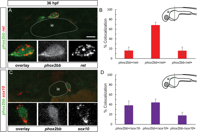
Enteric progenitor subpopulations are present at 36 hpf. A and C show confocal images of cross-sections through the trunk. (A) At 36 hpf, ret mRNA (red) partially colocalizes with phox2bb (green). Insets show close-up of outlined cell, overlay, phox2bb, ret mRNA (from left to right). Note that phox2bb refers to phox2bb:EGFP expression as described in the text. (B) Quantification in percent of phox2bb and ret colocalization at the wave front. (C) sox10 mRNA (red) partially colocalizes with phox2bb (green). Insets show close-up of outlined cell, overlay, phox2bb, sox10 mRNA (from left to right). (D) Quantification in percent of phox2bb and sox10 colocalization at the wave front. ie, intestinal epithelium Scale bar = 10 μm.