
|
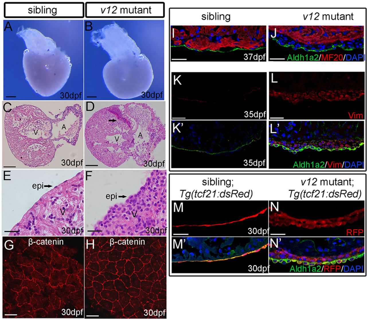
Fig. 3
Overproliferation of Tcf21-positive epicardial-derived cells in v12 mutants. (A,B) Ventricular morphology (dorsal view) of the v12 mutant and a wild-type sibling at 30 dpf. Note the spherical ventricles in the v12 mutant (B) as compared with the pyramid-like heart in wild type (A). (C-F) Histology of the v12 mutant (D,F) and its wild-type sibling (C,E), showing the malformed heart and sub-epicardial hemorrhage (arrow) in the mutant. Epicardial cells (epi) change from wild-type squamous epithelium (E) to oval-shaped in the v12 mutant (F). (G,H) β-catenin immunostaining marks the cell membrane of epicardial cells. Cell size is reduced and the cell boundary is smoother in the mutant (H) compared with the wild-type sibling (G). (I,J) MF20 staining shows that the overproliferated cells in the sub-epicardium of the v12 mutant are negative for muscle-specific myosin heavy chain. (K-L') The overproliferated cells are positive for vimentin (a marker of mesenchymally derived cells) in the v12 mutant (L,L') compared with its control sibling (K,K'). Aldhla2 marks the epicardium. (M-N') Aldh1a2 colocalizes with Tg(tcf21:dsRed) transgenic reporter in the epicardial cells of the wild-type sibling at 30 dpf (M,M'). By contrast, both overproliferated cells in the sub-epicardium region and the outermost layer of epicardial cells are positive for DsRed staining. Only the outermost layer of epicardial cells shows strong Aldh1a2 staining in the mutant (N,N'). A, atrium; V, ventricle. Scale bars: 100 μm in A-D; 20 μm in E-N.