
|
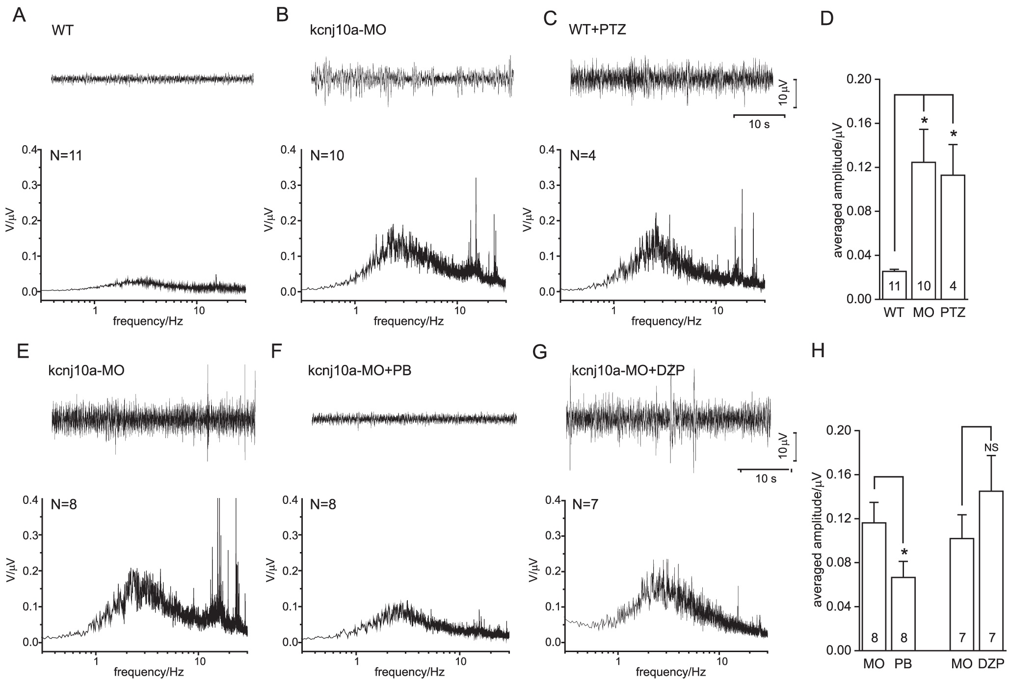
Fig. 3
Analysis of surface EEGs.
(A-C) Original traces (top) and averaged frequency spectra (below) obtained in WT ZF, kcnj10a morphant ZF and WT ZF pretreated with pentylenetetrazol. (D) The averaged amplitudes (mean over 2-4 Hz) from (A-C) are compared. Note the significant epileptic activity in kcnj10a morphant (MO) and pentylenetetrazol-treated (PTZ) ZF. (E-G) Original traces (top) and averaged frequency spectra (below) obtained in kcnj10a morphant fish, and kcnj10a morphants treated with pentobarbitone (PB) or diazepam (DZP), respectively. (H) Averaged amplitudes (mean over 2-4 Hz) from (E-G) are compared. Note the significant suppression of epileptic activity by PB treatment, and lack of suppression by DZP.