
Image
|
Figure Caption
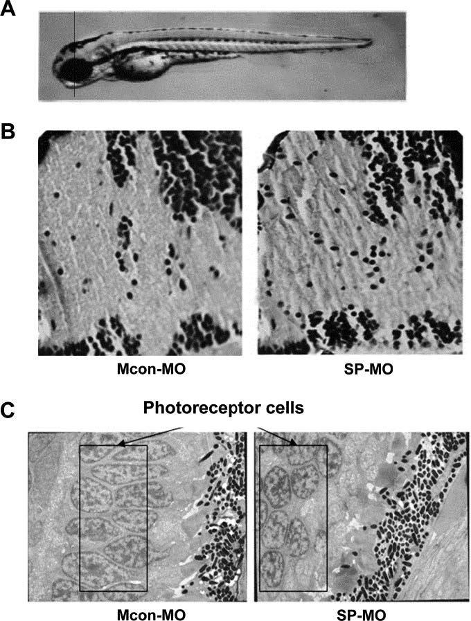
Fig. S6
Abnormities of ztfpi-2 knockdown morphants in nerve fibers and retina at 96 hpf. (A) Diagram showing the plane of section. (B) H&E-stained slides of the wild-type and mutant encephalon magnified 100×. Interspaces among the nerve fibers in ztfpi-2 morphants became wider than those in the control. (C) The arrangement of retina photoreceptor cells became disorganized when ztfpi-2 was downregulated by 0.5 mM SPMO (black rectangle).
Figure Data
Acknowledgments
This image is the copyrighted work of the attributed author or publisher, and
ZFIN has permission only to display this image to its users.
Additional permissions should be obtained from the applicable author or publisher of the image.
Reprinted from Developmental Biology, 381(1), Zhang, Y., Wang, L., Zhou, W., Wang, H., Zhang, J., Deng, S., Li, W., Li, H., Mao, Z., and Ma, D., Tissue factor pathway inhibitor-2: A novel gene involved in zebrafish central nervous system development, 38-49, Copyright (2013) with permission from Elsevier. Full text @ Dev. Biol.