
|
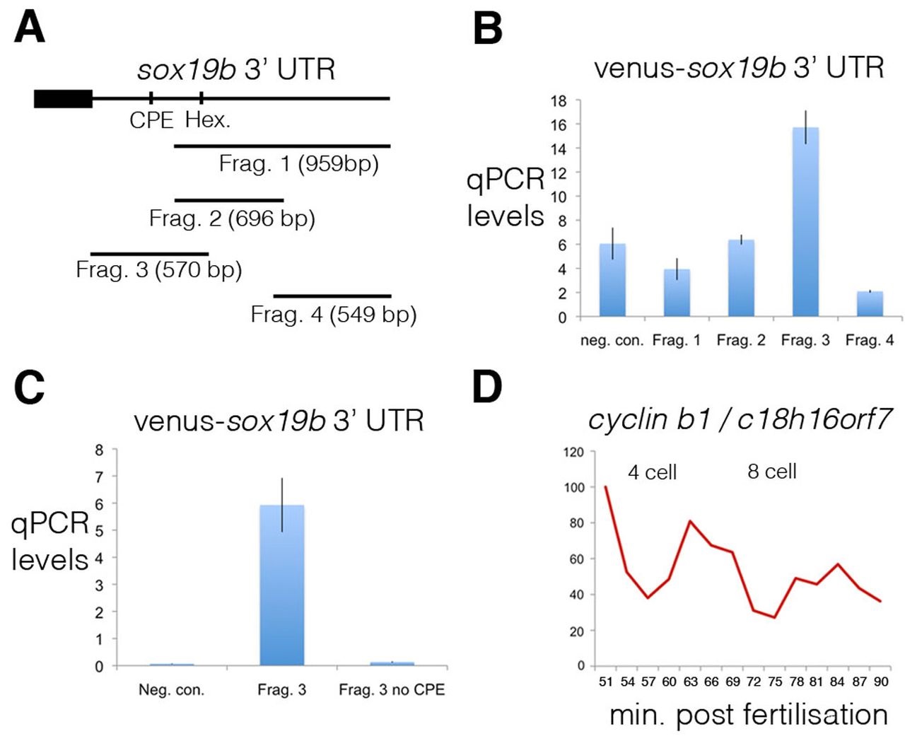
Fig. 4
Cytoplasmic polyadenylation elements drive the post-transcriptional regulation of maternal mRNAs. (A) To map the elements controlling polyadenylation of the maternal mRNA sox19b, four different fragments of the 3′ UTR were cloned downstream of the coding sequence of the fluorescent protein Venus. The positions of a cytoplasmic polyadenylation element (CPE) and hexamer, which were contained within fragment 3, are shown. (B) Injection of fertilised zebrafish embryos with Venus-sox19b 3′ UTR RNA demonstrated that only fragment 3 is polyadenylated. The ratio of quantitative PCR levels at the 64-cell stage to 2-cell stage are shown. cDNA was generated with oligo(dT) primers. As a negative control (neg. con.), embryos were injected with Venus RNA without a 3′ UTR. Error bars indicate s.d. (C) A CPE is present within fragment 3 of the sox19b 3′ UTR, which when deleted abolishes polyadenylation. (D) To study the cell cycle-dependent post-transcriptional regulation of cyclin B1, quantitative PCR was performed on cDNA, generated with oligo(dT) primers, derived from embryos synchronised by IVF. Embryos were collected every 3 minutes, starting just after the 2-cell stage. cyclin B1 polyadenylation levels are regulated in a cell cycle-dependent manner, with levels peaking during mitosis.