
Fig. S4
- ID
- ZDB-IMAGE-161102-20
- Genes
- Publication
- Lin et al., 2013 - MicroRNA-3906 Regulates Fast Muscle Differentiation through Modulating the Target Gene homer-1b in Zebrafish Embryos
- All Figures
- Figures for Lin et al., 2013
|
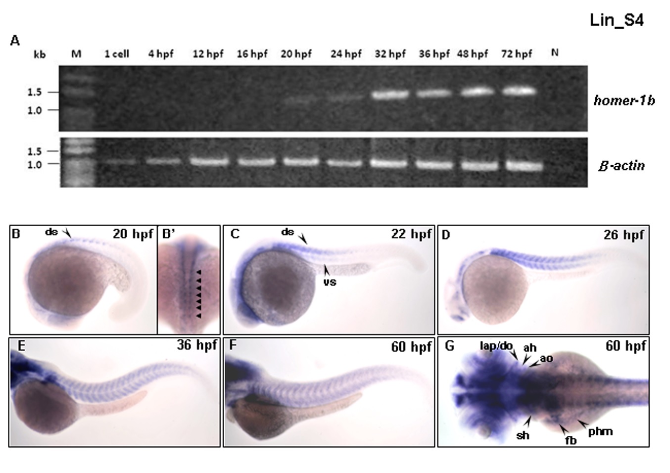
Fig. S4
The expression patterns of homer-1b mRNA at various developmental stages. (A) Using reverse transcription-PCR to detect homer-1b transcripts in embryos at various stages as indicated. The homer1b cDNA was detected from 20 hpf until at least 72 hpf. No homer-1b primers were added to serve as negative control. Detection of β-actin cDNA served a positive control. Using whole mount in situ hybridization to detect the expression patterns of homer-1b mRNA in the muscle region of zebrafish embryos at indicated stages. (B-F) were lateral view and (B′, G) were dorsal view. The homer-1b was starting to express at the dorsal somite (ds) in the front of mature somites at 20 hpf (B, B′, arrow). At 22 hpf, homer-1b was detected at dorsal region (dorsal somite, ds) and ventral region (ventral somite, vs) in the front of mature somites (C, arrow). At 26 hpf, homer-1b expression was increased in the newly formed somites in tail (D). At 36 hpf, homer-1b transcripts were detected in trunk muscles (E, F). (G) At 60 hpf, homer-1b was also expressed in the trunk migratory muscles of fin bud (fb), posterior hypoaxial muscle (phm) and sternohyoideus (sh), as well as craniofacial muscles of adductor hyoideus (ah), adductor operculi (ao), dilator operculi (do) and levator arcus palatini (lap).