
Fig. 7
- ID
- ZDB-IMAGE-161102-18
- Publication
- Lin et al., 2013 - MicroRNA-3906 Regulates Fast Muscle Differentiation through Modulating the Target Gene homer-1b in Zebrafish Embryos
- All Figures
- Figures for Lin et al., 2013
|
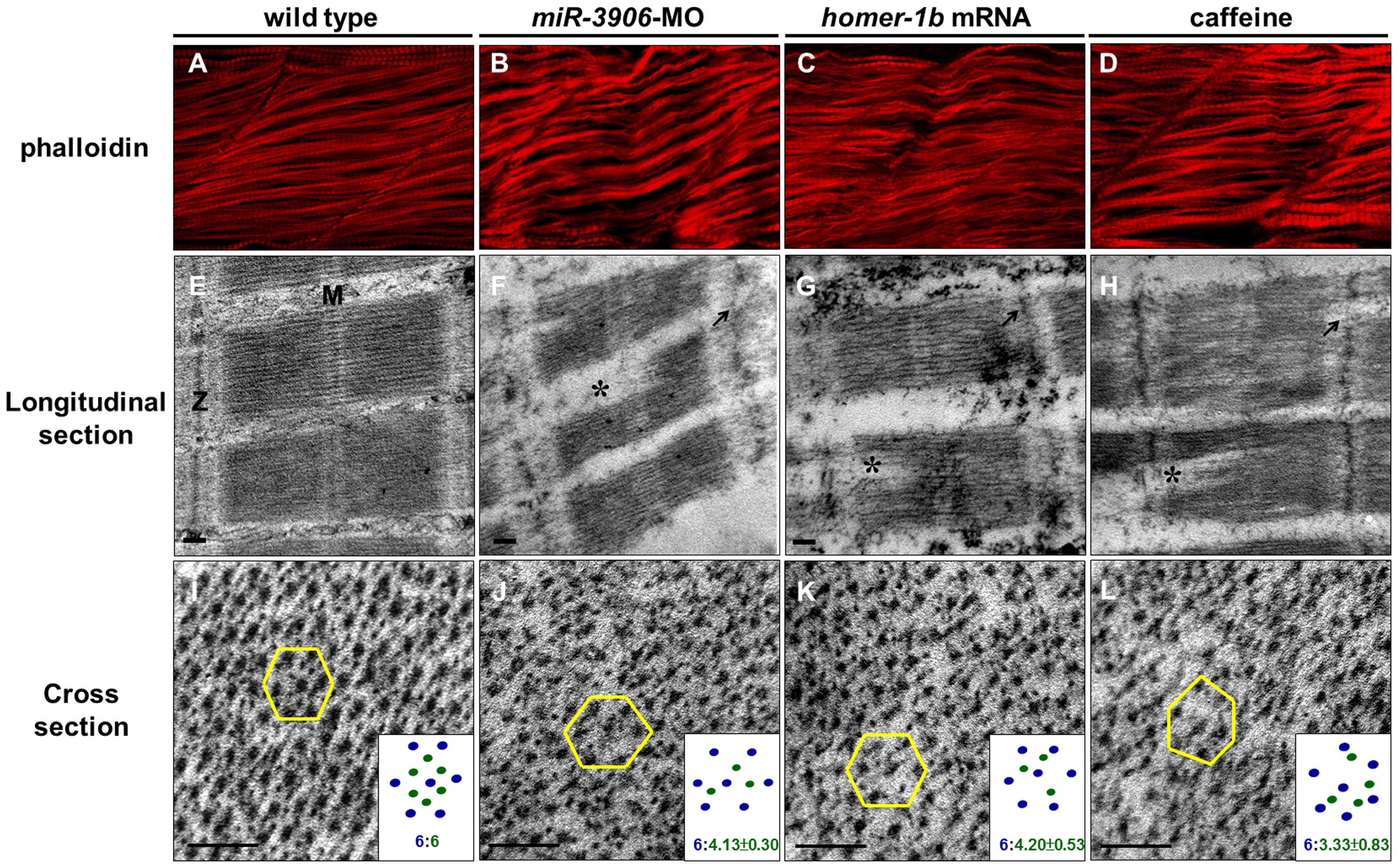
Fig. 7
Either inhibition of miR-3906 or overexpression of homer-1b disrupts sarcomeric actin organization.
Using phalloidin stain, compared to (A) WT embryos, (B) miR-3906-MO-injected embryos, (C) homer-1b-mRNA-injected embryos and (D) caffeine-soaked embryos exhibited a bending and disruption of sarcomeric actin organization in muscle cells. In longitudinal section, compared to (E) WT control embryos, (F) the Z-disc structure displaying in miR-3906-MO-injected embryos, (G) homer-1b-mRNA-injected embryos and (H) caffeine-soaked embryos was chaotic (indicated by arrows), and the full-length sarcomeres in muscle fibers were interrupted (indicated by asterisks) and could not be seen completely in a full sarcomere line. In cross section, compared to (I) WT control embryos, the relative position and proportion between myosin heavy chain and actin filament among the (J) miR-3906-MO-injected embryos, (K) homer-1b-mRNA-injected embryos and (L) caffeine-soaked embryos were chaotic and extremely irregular. For each sample, we randomly selected five positions to identify their hexagonal arrangements and calculated the ratio between the thick and thin filaments (n = 3). Results were presented at the bottom right of each picture (I-L). M:M line;Z:Z-disc;scale bar:0.1 µm.