
Fig. 2
- ID
- ZDB-IMAGE-161102-13
- Genes
- Publication
- Lin et al., 2013 - MicroRNA-3906 Regulates Fast Muscle Differentiation through Modulating the Target Gene homer-1b in Zebrafish Embryos
- All Figures
- Figures for Lin et al., 2013
|
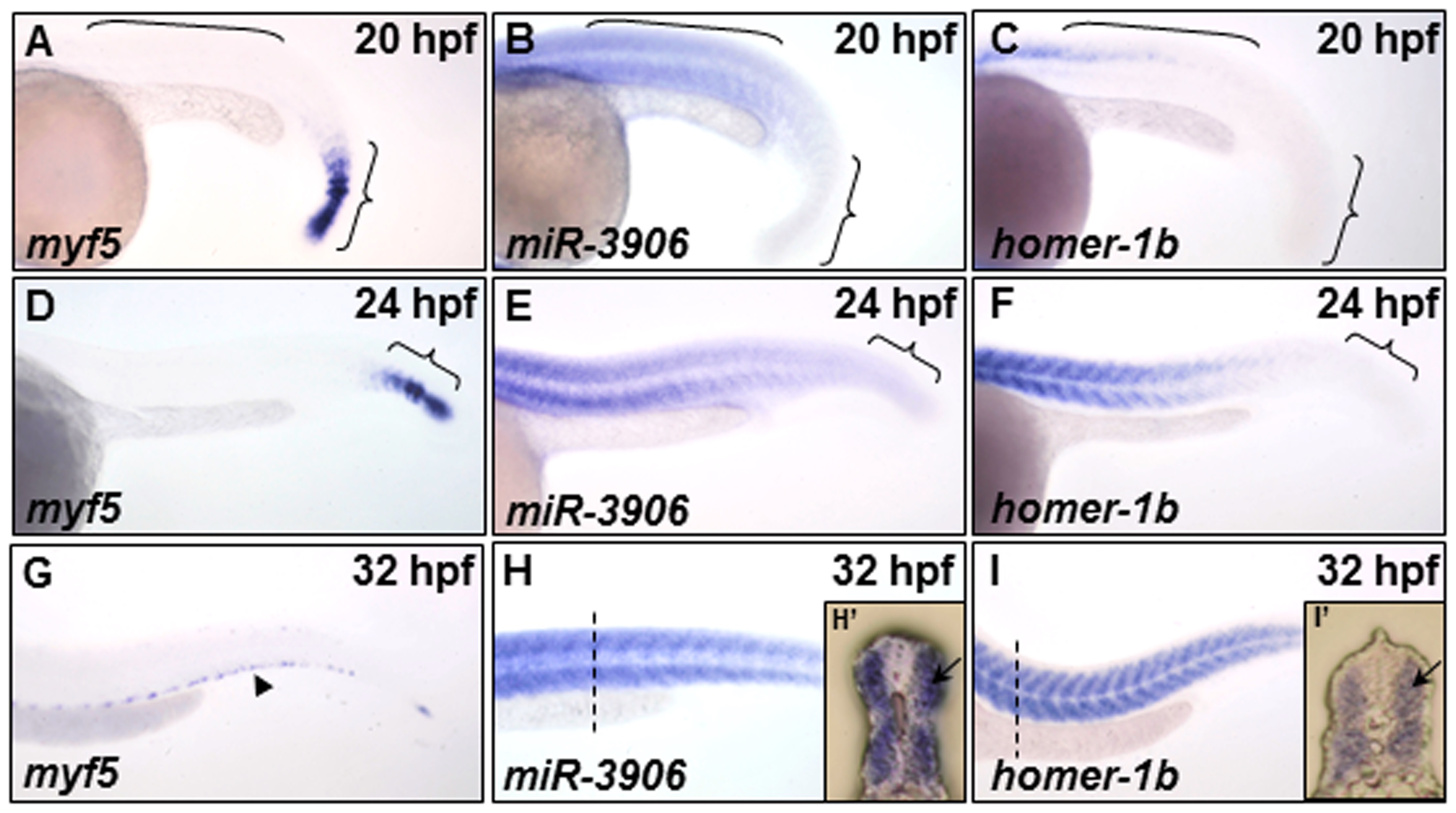
Fig. 2
The expression patterns of myf5, miR-3906 and homer-1b in the trunk somites of zebrafish embryos.
(A, D, G) Using whole-mount in situ hybridization to detect the temporal and spatial expression patterns of myf5, (B, E, H) miR-3906 and (C, F, I) homer-1b in zebrafish embryos at 20 hpf, 24 hpf and 32 hpf. At 20 hpf, (A) myf5 was expressed in the pre-somitic mesoderm and newly formed somites (indicated by { ), but it was absent in mature somites (indicated by [); (B) miR-3906 and (C) homer-1b were detected in mature somites. At 24 hpf, (D) myf5 was detected in the pre-somitic mesoderm, while (E) miR-3906 and (F) homer-1b gradually increased expression in mature somites. At 32 hpf, (G) myf5 was expressed in the edge region of trunk muscles (arrowhead), but (H) miR-3906 and (I) homer-1b were expressed in all trunk muscles. Cross section (dotted lines) showed that (F′) miR-3906 and (I′) homer-1b were expressed in fast muscle (arrows).