
|
Fig. S1
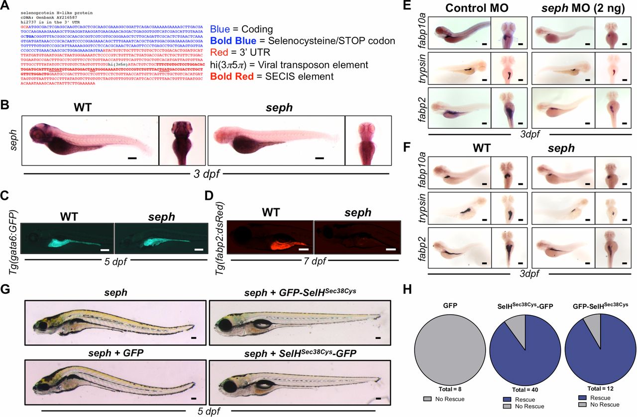
seph mutant Zebrafish exhibit defects in organ development. (A) Sequence of seph mutant reveals the viral insertion in the 3′ UTR that displaces the SECIS regulatory element that is required for selenoprotein synthesis. (B) WISH analysis of seph expression in WT and seph mutants at 3 dpf. (Scale bar: 200 µm.) (C) Fluorescent imaging of WT and seph mutant larvae on a Tg(gata6:GFP) background at 5 dpf. (Scale bar: 200 µm.) (D) Fluorescent imaging of WT and seph mutant larvae on a Tg(fabp2:dsRed) background at 7 dpf. (Scale bar: 200 µm.) (E) WISH analysis of fabp10a (liver), trypsin (pancreas), and fabp2 (intestine) expression in WT and seph morphants (2 ng, ATG morpholino) at 3 dpf. (Scale bar: 200 µm.) (F) WISH of fabp10a (liver), trypsin (pancreas), and fabp2 (intestine) expression in WT and seph mutants at 3 dpf. (Scale bar: 200 µm.) (G) Morphological assessment illustrating the rescue of the seph mutant phenotype by the injection of SelH-GFP mRNA (GFP-SelHSec38Cys and SelHSec38Cys-GFP constructs) at the one-cell stage. (H) Quantitative determination of the rescue of the seph mutant phenotype by the injection of SelH-GFP mRNA (GFP-SelHSec38Cys and SelHSec38Cys-GFP constructs) at the one-cell stage.