
|
Fig. 6
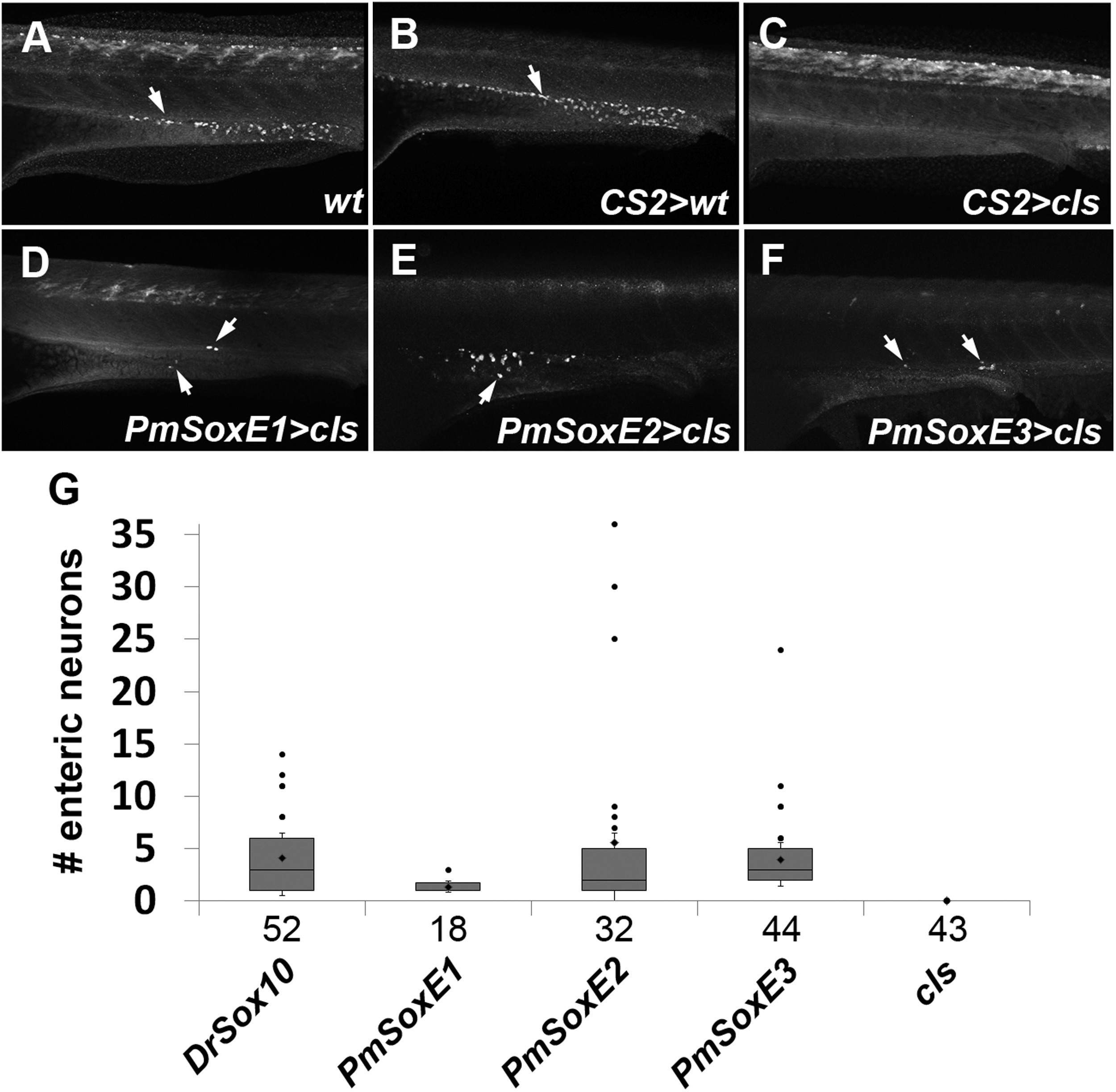
pCS2-PmSoxE heterospecific expression promotes differentiation of enteric neurons along the larval gut in cls mutants. (A-F) anti-HuC/D immunofluorescence in 96 hpf zebrafish. (A) wildtype. (B) control mock-injected wildtype. (C) cls embryo mock-injected with empty pCS2 vector. Note the absence of HuC/D-positive enteric neurons in cls embryos. Enteric neurons are present in cls mutants injected with lamprey PmSoxE1 (D), PmSoxE2 (E), and PmSoxE3 (F) constructs (arrows in D-F). (G) box and whisker plots indicate the number of enteric neurons present in zebrafish injected with Drsox10, PmSoxE1, PmSoxE2, and PmSoxE3 constructs. Whiskers represent standard error. Each dot above the positive whiskers represents individual data points of the first quartile. Sample sizes are indicated under each bar on the X-axis. Arrows indicate anti-HuC/D positive enteric neurons. Orientation: anterior facing left.
Reprinted from Developmental Biology, 418(1), Lee, E.M., Yuan, T., Ballim, R.D., Nguyen, K., Kelsh, R.N., Medeiros, D.M., McCauley, D.W., Functional constraints on SoxE proteins in neural crest development: the importance of differential expression for evolution of protein activity, 166-78, Copyright (2016) with permission from Elsevier. Full text @ Dev. Biol.