
Image
|
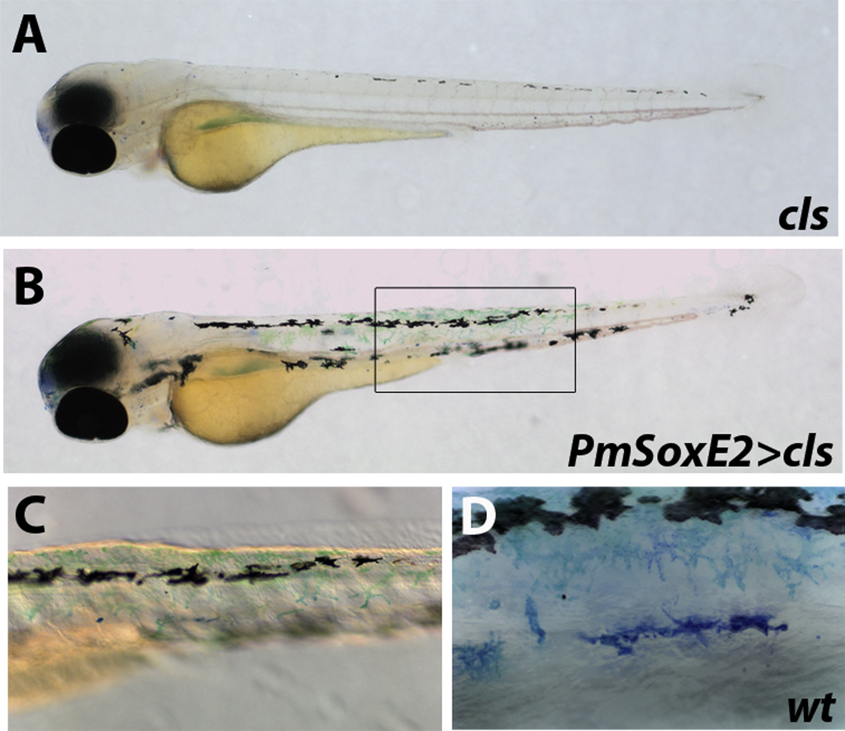
Figure Caption
Fig. 5
PmSoxE2 promotes differentiation of xanthophores in cls mutant zebrafish. (A-B) 76 hpf zebrafish embryos stained with methylene blue to detect presence of xanthophores. (A) cls mutant lacking differentiated xanthophores. (B) xanthophores in cls mutants injected with the pCS2-PmSoxE2 construct. (C) inset in “B” ; higher magnification highlights the presence of xanthophores following PmSoxE2 injection. (D) morphology of xanthophores in a wildtype 76 hpf embryo. Orientation: anterior facing left.
Acknowledgments
This image is the copyrighted work of the attributed author or publisher, and
ZFIN has permission only to display this image to its users.
Additional permissions should be obtained from the applicable author or publisher of the image.
Reprinted from Developmental Biology, 418(1), Lee, E.M., Yuan, T., Ballim, R.D., Nguyen, K., Kelsh, R.N., Medeiros, D.M., McCauley, D.W., Functional constraints on SoxE proteins in neural crest development: the importance of differential expression for evolution of protein activity, 166-78, Copyright (2016) with permission from Elsevier. Full text @ Dev. Biol.