
|
Fig. 3
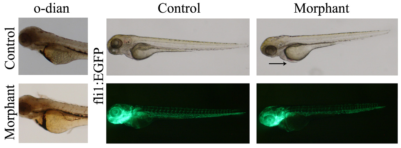
Blood pooling in morphant embryos. (A,B) Lateral view of O-dianisidine stained 72 hpf (A) control and (B) morpholino injected embryos. Blood was present in the anterior blood vessels, the heart, the common cardinal vein, the dorsal aorta, and posterior cardinal vein. In the morphant embryos, blood was pooled on the yolk prior to entering the heart. (C–F) The endothelial specific transgenic fli1:EGFP embryos injected with (E) control or (F) plakophilin 2 morpholino were analysed under light (C and D) and fluorescence microscopy (E and F) for vascular defects at 72 hpf. There was no alteration in vasculature in morphant embryos despite evident blood pooling (arrow).