
|
Fig. S4
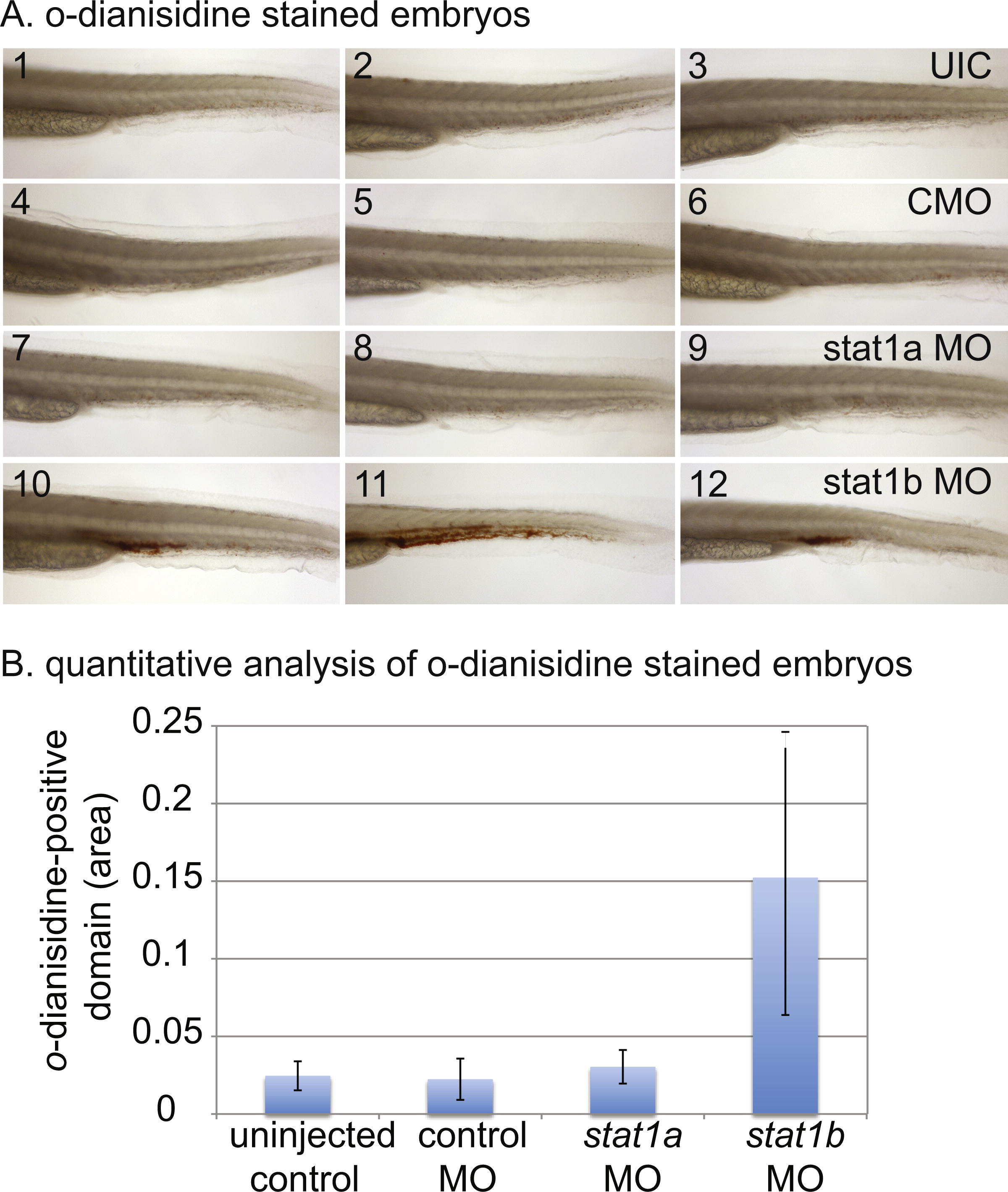
Histochemical staining of hemoglobin with o-dianisidine. A. Embryos stained with o-dianisidine: uninjected controls (UIC, 1-3), control MO injected embryos (CMO, 4-6), stat1a MO treated embryos (7-9), and stat1b MO treated embryos (10-12). Knockdown of stat1b caused an increase in hemoglobin staining in the trunk compared to controls and stat1a MO injected embryos. B. Quantitative analysis of o-dianisidine-stained embryos, ten individuals scored for each of the four conditions. Pixels of o-dianisidine positive cells were counted and summed. Compared to the uninjected control using a t-test (single tailed), P values for significant only for stat1b MO (p = 0.0014), and were not significant for control MO (0.337) or stat1a MO (0.108).
Reprinted from Mechanisms of Development, 128(7-10), Song, H., Yan, Y.L., Titus, T., He, X., and Postlethwait, J.H., The role of stat1b in zebrafish hematopoiesis, 442-56, Copyright (2011) with permission from Elsevier. Full text @ Mech. Dev.