
Fig. 1
- ID
- ZDB-IMAGE-161020-5
- Publication
- Granato et al., 1996 - Genes controlling and mediating locomotion behavior of the zebrafish embryo and larva
- All Figures
- Figures for Granato et al., 1996
|
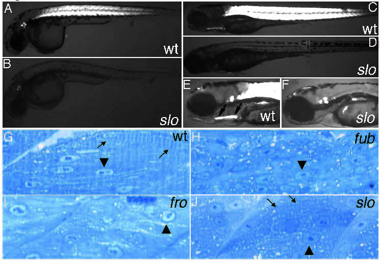
Fig. 1
Comparison of muscle differentiation between wild-type (A,C,E,G), slo (B,D,F,J), fro (I) and fub (H) embryos using polarized light (A-F) or trunk muscle sections (G-J). At 30 hours birefringency is reduced in slo embryos (B) compared to wild-type embryos (A). At later stages (96 hours) muscle birefringency is detectable in somitic muscle and in jaw muscle of wild-type larvae (C, E), while in slo mutants (D, F) birefringency is completely absent in somitic and jaw muscles. The arrows in E delineate the jaw muscles. (G) Sagitallateral section through 36 hours wild-type somitic muscle tissue showing muscle fibers (arrows) and elongated nuclei (arrowhead). In fub mutants muscle fibers are absent; however, the nuclei are elongated (arrowhead) and resemble those of wild type (H). In contrast, nuclei in fro (I) and slo (J) are round (arrowhead). (J) Occasionally muscle fibers can be detected in slo somitic tissue (arrows).