
|
Fig. 4
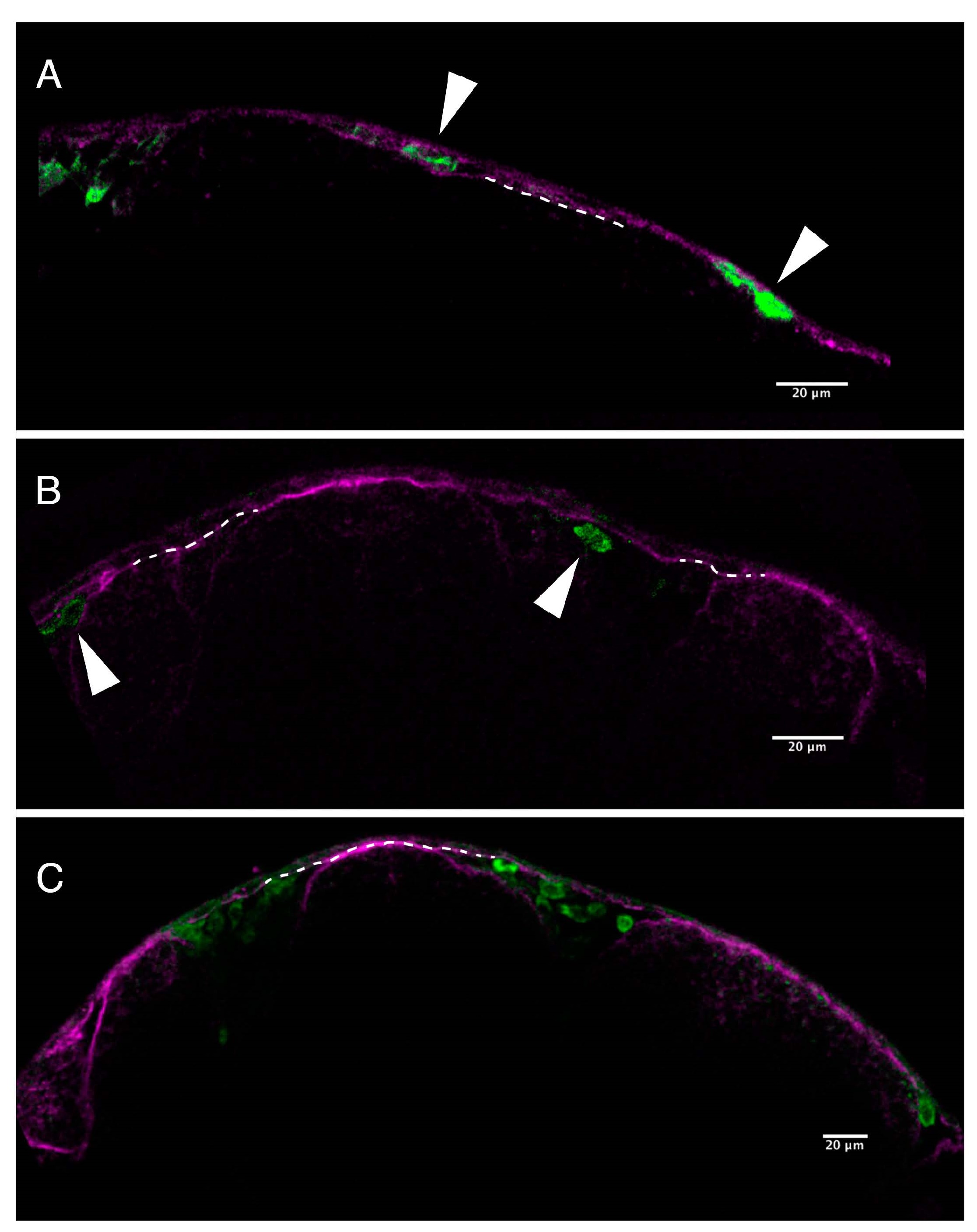
Pigment cells fail to invade epidermis in the presence of PhN. (A) A single focal plane through the head epidermis of a Tg(mitfa:eGFP) embryo exposed from 18 to 24 hpf to 10 µM PhE and stained with anti-laminin (magenta) and anti-GFP (green) showing presumptive pigment cells that have crossed the basal lamina and successfully invaded the epidermis (arrowheads); (B) A comparable single focal plane through the head epidermis of a Tg(mitfa:eGFP) embryo exposed from 18 to 24 hpf to 10 µM PhN, showing presumptive pigment cells (arrowheads) having failed to invade across the basal lamina of the epidermis; (C) A slightly lower magnification view of a Tg(mitfa:eGFP) embryo exposed from 18 to 24 hpf to 10 µM PhN, showing several presumptive pigment cells trapped below the epidermis. Dotted lines indicate the basal lamina. Scale bars are 20 µm.