
|
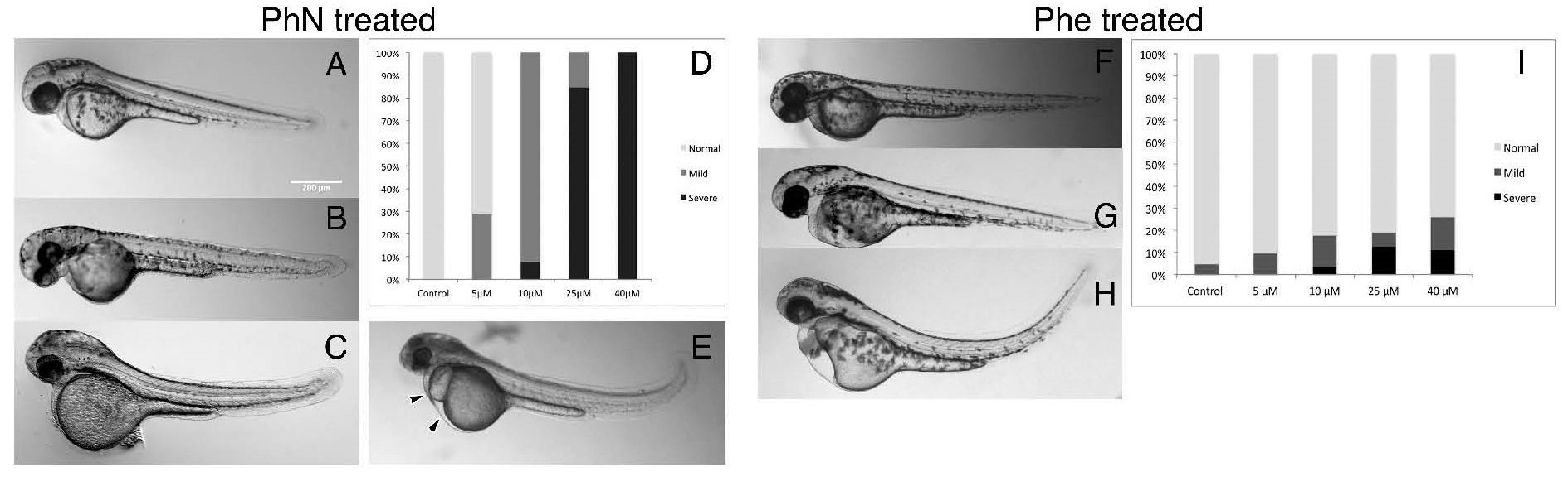
Fig. 2
PhN and PhE both disrupt normal development, but embryos are more sensitive to PhN. Embryos were treated with either PhN or PhE at various concentrations from 24 to 48 hours post-fertilization (hpf), and assessed (blinded) for gross morphological defects visually. (A) A normal embryo from the vehicle control group. Embryos treated with PhN representing individuals scored as: (B) "Normal"; (C) "Mild"; and (E) "Severe" phenotypes (pericardial and yolk-sac edema indicated with arrowheads); (D) Summary of results from scoring 44 embryos exposed to vehicle only, 69 exposed to 5 µM PhN, 63 exposed to 10 µM PhN, 62 exposed to 25 µM PhN, and 88 exposed to 40 µM PhN. Embryos exposed to PhE exhibiting: (F) "Normal"; (G) "Mild"; and (H) "Severe" phenotypes; (I) Summary of results from scoring of 60 embryos exposed to vehicle only, 48 exposed to 5 µM PhE, 47 exposed to 10 µM PhE, 51 exposed to 25 µM PhE, and 40 exposed to 40 µM PhE. In addition to tail curvature, necrosis, pericardial and yolk-sac edema seen in embryos exposed to either PhE or PhN, embryos exposed to PhN exhibited loss of pigmentation, craniofacial abnormalities, and absence of intersomitic circulation. Scale bar = 200 µm.