
Image
|
Figure Caption
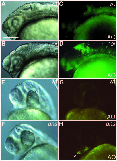
Fig. 6
Mutants that show abnormaly localized apoptotic cells. (A,C,E,G) Wild type; (B,D) noi; (F,H) dns. (A-D) At 24 h; (E-H) at 36 hours. (A-D) Localized apoptotic cells are seen in the presumptive isthmus in noi (arrowhead); (E-H) restricted apoptotic cells are detected in the anterior prechodal plate in dns embryos (arrowhead). Scale bar, 100 µm.
Figure Data
Acknowledgments
This image is the copyrighted work of the attributed author or publisher, and
ZFIN has permission only to display this image to its users.
Additional permissions should be obtained from the applicable author or publisher of the image.
Full text @ Development