
|
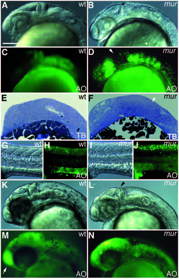
Fig. 3
Class II mutant phenotypes. (A,C,E,G,H,K,M) Wild type embryos; (B,D,F,I,J,L,N) mur embryos. (A-J) at 24 hours, (K-N) at 36 hours. (C,D,H,J,M,N) Acridine orange (AO)-stained embryos of A,B,G,I,K,L, respectively. (E,F) Histological structure of the brain, as shown in parasagittal section. (A,B,K,L) Pointed midbrainhindbrain boundary (black arrowhead) is seen in mur embryos; (C,D) apoptotic cells are seen in parts of the forebrain, hindbrain and the spinal cord (H,J). The midbrain (white arrowhead in D) is less affected. (E,F) Toluidine blue stained apoptotic neuroepithelium is not seen in the midbrain (white arrowhead) in mur embryos; (K,L) small and less pigmented head of mur embryos; (M,N) apoptotic cells are seen in dorsal diencephalon and midbrain. Apoptotic cells in the hindbrain and the spinal cord are reduced. Acridine orange stained olfactory epithelium (white arrow in M) is reduced in mur embryos. Scale bar, 100 µm.