
|
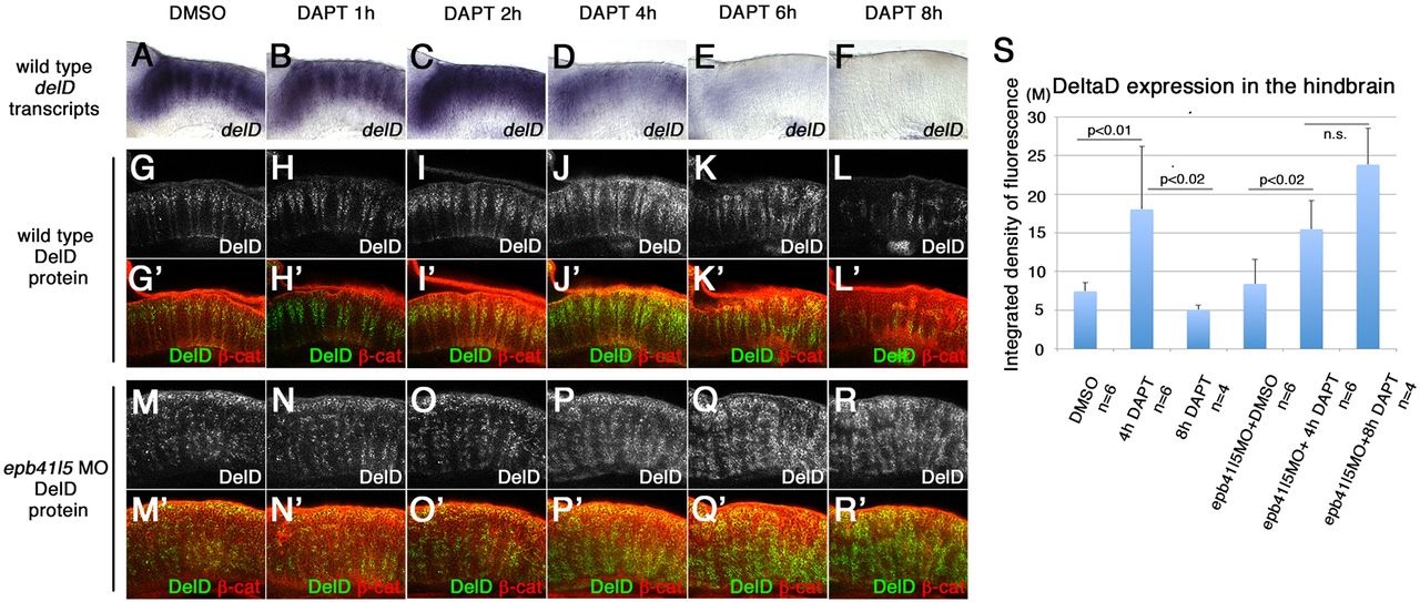
Fig. 4
‘Intermediate’ neuronal progenitor population expressing DeltaD is increased in epb41l5-deficient embryos. (A-L′) Timeline of disappearance of DeltaD in progenitors during neuronal differentiation in wild-type embryos. Two hours of DAPT treatment increases expression of deltaD transcripts, followed by increased expression of DeltaD protein at 4h. Expression of deltaD and DeltaD diminishes after 6 and 8h of DAPT treatment. (M-R′) Continued expression of DeltaD in epb41l5 morphants. Increased DeltaD expression persists after 8h of DAPT treatment. (S) Quantitative analysis of DeltaD expression. Total fluorescence intensity of DeltaD in the hindbrain was measured in individual confocal slices using ImageJ. Error bars represent s.d. n.s., not significant. M, million integrated pixel intensity.