
Image
|
Figure Caption
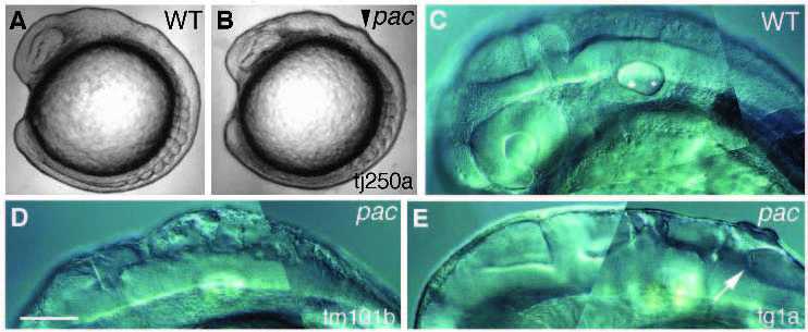
Fig. 6
Phenotype of live pac embryo. C-E treated with 0.2 mM PTU. Lateral views of wild-type (A) and pactj250a (B) embryos at around the 9-somite stage. The small bulge (arrowhead) in the hindbrain is indicated. Lateral views of wild type (C) at 28 hpf, pactm101b (D) at 28 hpf and pactg1a (E) at 36 hpf. The filopodia-like structure (arrow) is indicated. Bar, 320 µm (A,B); 200 µm (C-F).
Figure Data
Acknowledgments
This image is the copyrighted work of the attributed author or publisher, and
ZFIN has permission only to display this image to its users.
Additional permissions should be obtained from the applicable author or publisher of the image.
Full text @ Development