
|
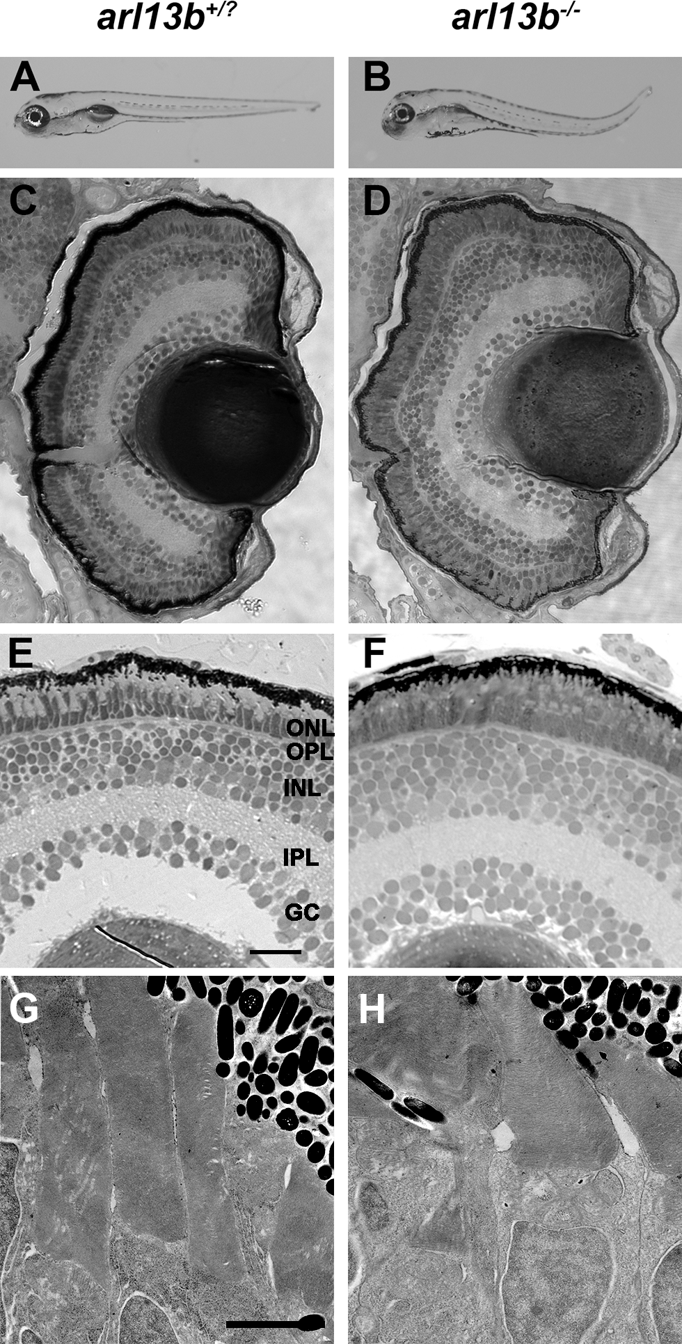
Fig. 1
arl13b is not required for zebrafish retinal development. (A, B) Lateral view of larvae at 5 dpf. (C-F) Methylene blue-stained plastic sections of 5 dpf wild-type siblings (arl13b+/?) and arl13b-/- mutant retinas. The outer nuclear layer (ONL), outer plexiform layer (OPL), inner nuclear layer (INL), inner plexiform layer (IPL), and ganglion cell layer (GC) were present in wild-type and mutant retinas. (G, H) Transmission electron micrographs of 5 dpf retinas do not show any evidence of photoreceptor outer segment disorganization or ciliary defects. Scale bars: 200 µm (A, B), 20 µm (C, D), 10 µm (E, F), and 2 µm (G, H).