
Image
|
Figure Caption
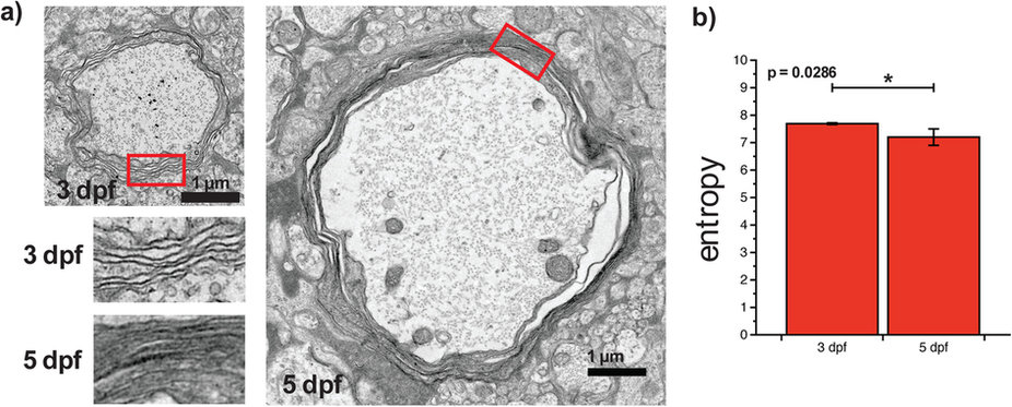
Fig. 4
Electron microscopy confirms that P-CARS informs on the nanostructure.
(a) Electron microscopy images at 3 and 5 dpf show the difference in myelin lamellae organization. (b) The entropy decreases significantly between 3 and 5 dpf (Mann-Whitney test, n = 4).
Acknowledgments
This image is the copyrighted work of the attributed author or publisher, and
ZFIN has permission only to display this image to its users.
Additional permissions should be obtained from the applicable author or publisher of the image.
Full text @ Sci. Rep.