
Image
|
Figure Caption
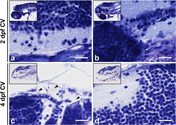
Fig. 6
Histopathological analysis of Streptococcus pneumoniae-infected zebrafish embryos via the caudal vein at 2 days post - fertilization. a, b Caudal vein injection at 2 days post - fertilization (dpf). Sagittal section of the head region showing the bacteria (arrows) in the brain parenchyma at a 12 h post injection (hpi) and at b 24 hpi. c, d Caudal vein-injected zebrafish embryos at 4 dpf. c Sagittal section at 24 hpi showing bacteria (arrows) in the meningeal space and in d the brain parenchyma. Scale bars, 10 µm
Acknowledgments
This image is the copyrighted work of the attributed author or publisher, and
ZFIN has permission only to display this image to its users.
Additional permissions should be obtained from the applicable author or publisher of the image.
Full text @ J Neuroinflammation