
Image
|
Figure Caption
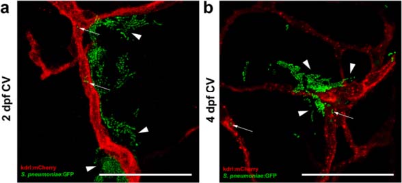
Fig. 5
Pneumococci leave the blood vessels after systemic infection. a, b Confocal microscopy images at maximum projection of Tg(kdrl:mcherry) s896 zebrafish embryos injected in the caudal vein (CV) a before formation of the blood-brain barrier (BBB) at 2 days post fertilization (dpf) or b after the formation of the BBB at 4 dpf. Bacteria were localized inside (arrows) and outside (arrow heads) the blood vessels. All embryos were infected with 400 CFU and imaged at 24 h post injection. Scale bars, 50 µm
Figure Data
Acknowledgments
This image is the copyrighted work of the attributed author or publisher, and
ZFIN has permission only to display this image to its users.
Additional permissions should be obtained from the applicable author or publisher of the image.
Full text @ J Neuroinflammation