
Image
|
Figure Caption
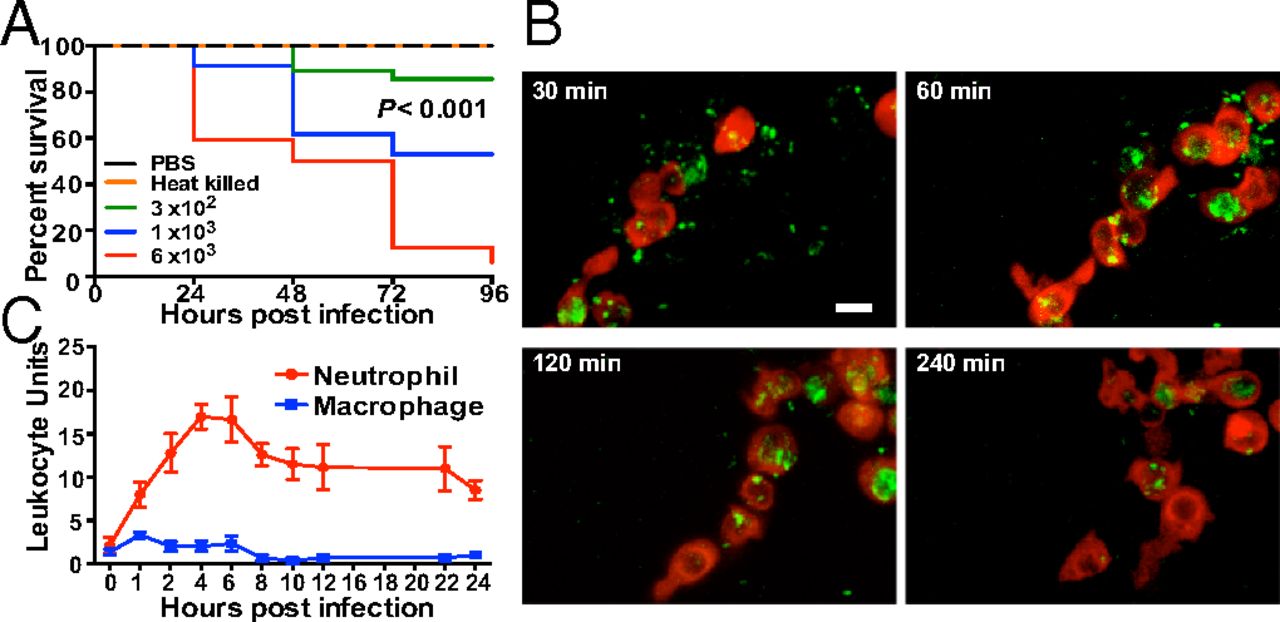
Fig. 1
A. baumannii infection in zebrafish. (A) Survival after bloodstream infection with live A. baumannii (n = 30 embryos, three biological replicates; P value is a comparison of 3 × 10² and 1 × 10³ cfu per embryo by log-rank test). (B) Time-lapse confocal scanning laser microscopy showing red neutrophils [Tg(lyz:DsRed)nz50] phagocytosing and clearing A. baumannii–GFP. (Scale bar: 20 µm.) (C) LUs after infection into the somatic muscle of zebrafish after adjustment for trauma (PBS solution injection; mean ± SEM, n = 5 per experiment, three biological replicates).
Figure Data
Acknowledgments
This image is the copyrighted work of the attributed author or publisher, and
ZFIN has permission only to display this image to its users.
Additional permissions should be obtained from the applicable author or publisher of the image.
Full text @ Proc. Natl. Acad. Sci. USA