
|
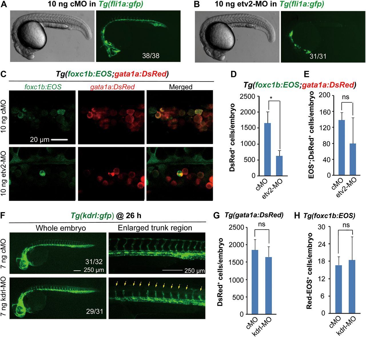
Fig. 4
Vasculature imperfection has no impact on hematopoiesis. (A and B) Effect of etv2 knockdown on vasculature formation. Note that etv2 morphants had normal morphology but lacked both dorsal aorta and cardinal vein in the trunk region. The ratio of embryos with the representative pattern was indicated. cMO, control morpholino. (C) foxc1b:EOS;gata1a:DsRed double-positive cells in the ICM of etv2 morphant or control at the 28s stage. (D and E) Sorting results of gata1a:DsRed+ (D) and fox1b:EOS+;gata1a:DsRed+ blood cells (E) in etv2 and control morphants at 30 hpf. The fluorescent cell number per embryo was averaged from three independent experiments (10 embryos each). *P < 0.05; ns, non-significance (P > 0.05). (F) kdrl knockdown impaired growth of intersegmental vessels (indicated by arrows). The ratio of affected embryos was indicated. (G and H) Effect of kdrl knockdown on LPM hematopoiesis (G) and somite hematopoiesis (H). For the injected Tg(foxc1b:EOS) embryos, five pairs of new born somites were photoactivated at the 20s stage. All embryonic cells were collected from a group of five embryos at 30 hpf and were sorted by flow cytometry for DsRed+ (G) or red-EOS+ cells (H). The number of cells with expected fluorescence per embryo was averaged from three independent experiments. ns, non-significance (P > 0.05).