
|
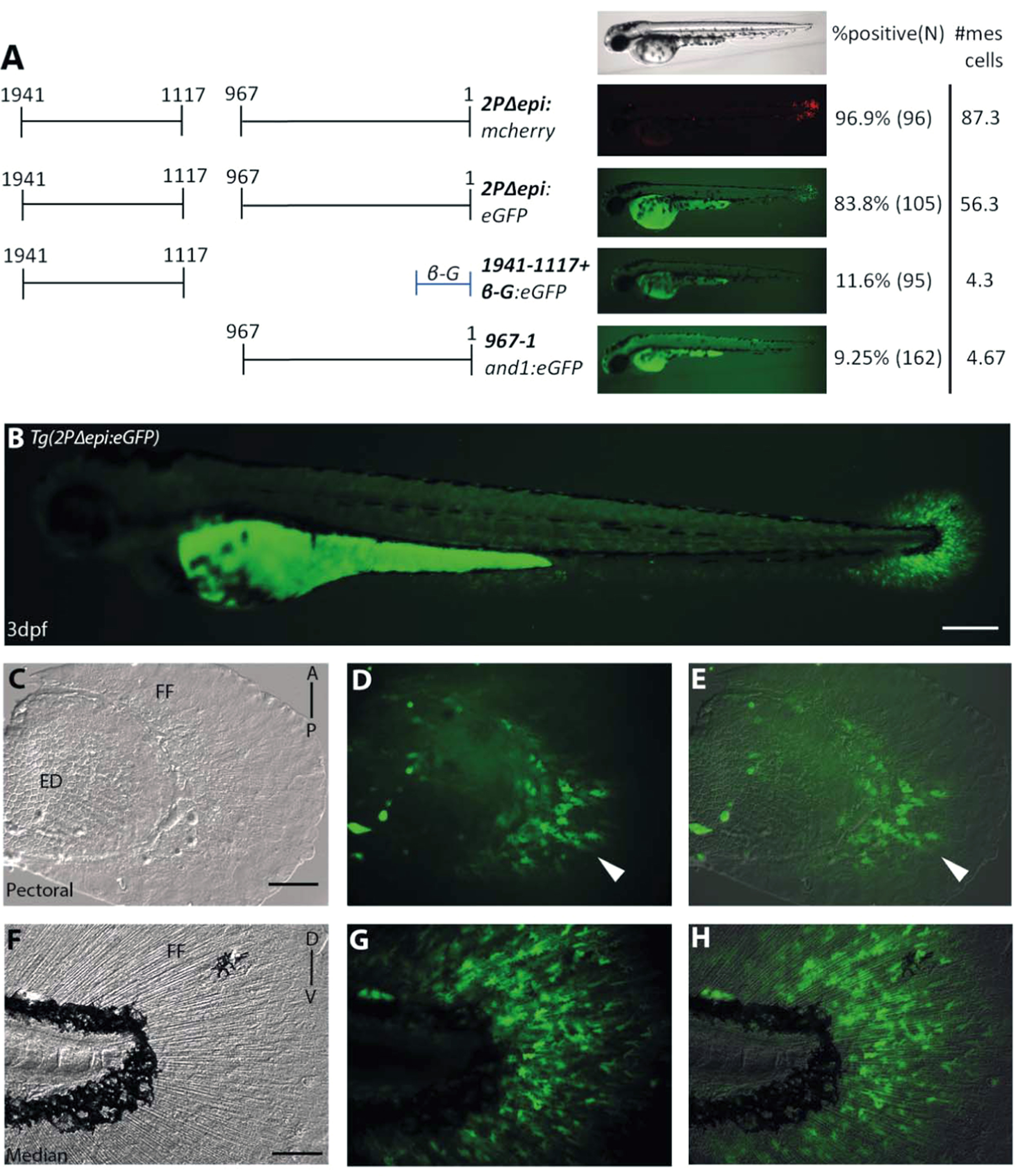
Fig. 5
Multiple mesenchymal enhancers exist and are required simultaneously to recapitulate and1 expression in mesenchymal cells of embryonic fin folds. (A) Analysis of truncated 2Pand1 reporter constructs. Constructs were tested for mesenchymal enhancer activity. Percentage of eGFP positive embryos during injections, and an average number of mesenchymal cells are presented on the right (N=Number of total viable injected embryos). Note the reduced percentages with Tg(1941-1117+β-globin:eGFP) and Tg(967and1:eGFP) constructs suggesting both fragments are required for full mesenchymal expression. (B-H) Tg(2PΔepi:eGFP) transgenic reporter line at 3 d.p.f. Median (F-H) and pectoral fin fold (C-E) display reporter expression in the mesenchymal cells only using the 1941-1117and1 and 967and1 fragments simultaneously. Note the higher number of eGFP-positive cells in the posterior half of the pectoral fin (D), indicated by the white arrowhead. Brightfield (C, F), fluorescence (B, D, G), and brightfield/fluorescence merged images (E, H). ED, Endoskeletal Disc; FF, Fin Fold; A, Anterior; P, Posterior; D, Dorsal; V, Ventral. Scale bars: 200 µm in B; 50 µm in C-H.
Reprinted from Developmental Biology, 417(1), Lalonde, R.L., Moses, D., Zhang, J., Cornell, N., Ekker, M., Akimenko, M.A., Differential actinodin1 regulation in zebrafish and mouse appendages, 91-103, Copyright (2016) with permission from Elsevier. Full text @ Dev. Biol.