
|
Fig. S8
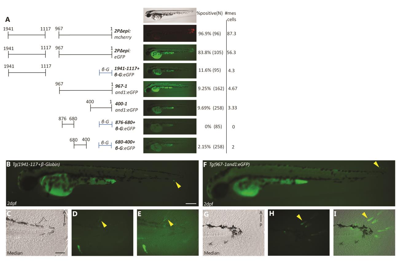
Full analysis for mesenchymal enhancer activity: multiple mesenchymal enhancers exist and are required simultaneously to recapitulate and1 mesenchymal cells in embryonic fin folds. (A) Transient expression analysis of truncated 2Pand1 reporter constructs in injected embryos. Constructs were tested for mesenchymal enhancer activity. Percentage of GFP positive embryos during injections, and an average number of mesenchymal cells are presented on the right (N = Number of total viable injected embryos). Note the reduced percentages of GFP positive embryos with the Tg(1941-1117+ β-globin:eGFP) and Tg(967-1and1:eGFP) constructs suggesting that both fragments are required for full mesenchymal expression (A). Note the even more reduced percentages obtained with smaller fragments Tg(400-1and1:eGFP), Tg(876-680+ β-globin:eGFP), Tg(680-400+β-globin:eGFP) (A). (B-E) Transient expression of Tg(1941-1117and1:eGFP) at 2 d.p.f. (F-I) Transient expression of Tg(967-1and1:eGFP) at 2 d.p.f. Neither fragment is capable of mimicking the expression of Tg(2PΔepi:eGFP). Note the very small amount of reporter expressing mesenchymal cells (indicated by yellow arrowheads) (B, D-E, F, H-I). Brightfield (C, G), fluorescence (B, D, F, H), and brightfield/fluorescence merged images (E, I). A, Anterior; P, Posterior; D, Dorsal; V, Ventral. Scale bars: 200µm in A-E; 50µm in B-D, F-H.
Reprinted from Developmental Biology, 417(1), Lalonde, R.L., Moses, D., Zhang, J., Cornell, N., Ekker, M., Akimenko, M.A., Differential actinodin1 regulation in zebrafish and mouse appendages, 91-103, Copyright (2016) with permission from Elsevier. Full text @ Dev. Biol.