
|
Fig. S7
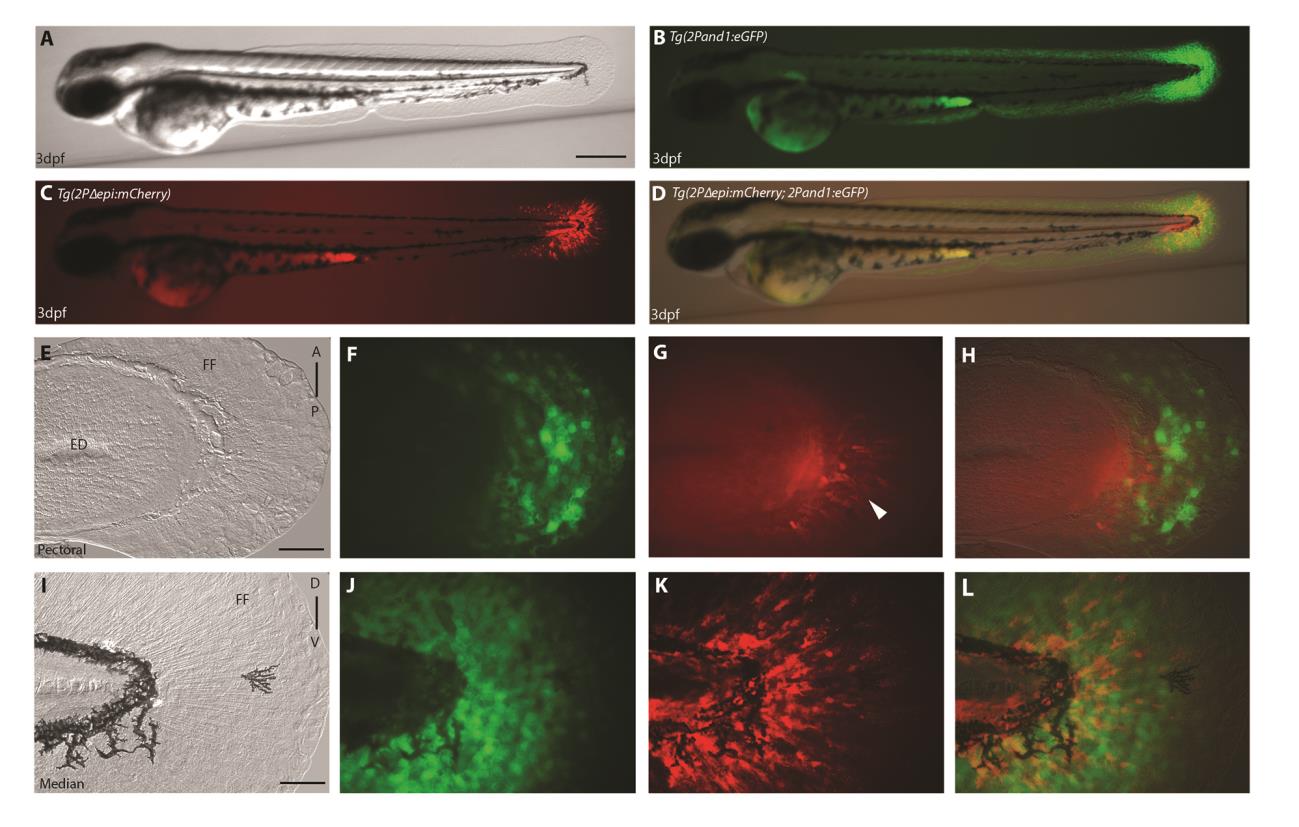
Reporter expression present in ectodermal and mesenchymal tissues in 3 d.p.f median and pectoral fin folds. (A-L) Double transgenic reporter line Tg(2Pand1:eGFP; 2PΔepi:mCherry) at 3 d.p.f., allowing visualization of the ectodermal- and mesenchymal-expressing cells simultaneously. Despite the presence of mesenchymal expression within Tg(2Pand1:eGFP), this line was used to assess ectodermal reporter expression to ensure all potential cis-regulatory elements are included. Tg(2Pand1:eGFP) reporter expression in ectodermal cells and mesenchymal cells are presented in green (B, F, J), Tg(2PΔepi:mCherry) reporter expression in mesenchymal cells are presented in red (C, G, K). Ectodermal and mesenchymal images are merged (D, H, L). Ectodermal and mesenchymal images are merged (D, H, L) to visualize ectodermal eGFP and mesenchymal mCherry expression simultaneously. By 3 d.p.f, ectodermal and mesenchymal expression is present in the median and pectoral fin fold (A-L). Note the stronger mesenchymal expression in the posterior half of the pectoral fin fold (indicated by the white arrowhead) (G). Tip of the yolk sac extension displays autofluorescence (B-D). ED, Endoskeletal Disc; FF, Fin Fold; A, Anterior; P, Posterior; D, Dorsal; V, Ventral. Scale bars: 300µm in A-D, 50µm in E-L.
Reprinted from Developmental Biology, 417(1), Lalonde, R.L., Moses, D., Zhang, J., Cornell, N., Ekker, M., Akimenko, M.A., Differential actinodin1 regulation in zebrafish and mouse appendages, 91-103, Copyright (2016) with permission from Elsevier. Full text @ Dev. Biol.